On September 25–27, the biodrook team joined IT Arena 2026 in Lviv one of Ukraine’s leading technology events, bringing together innovators, founders, investors, and technology leaders from Ukraine and around the world. This year’s conversations covered some of the topics especially relevant to us as a growing medtech company: AI, product development, investment, scaling, emerging technologies, and building Ukrainian companies for global markets. IT Arena was a valuable opportunity to look beyond the medtech field, learn how other technology companies approach growth and product development, exchange ideas, and connect with people building the next generation of Ukrainian innovation. One of our key takeaways: a strong technology is only the beginning. Turning it into real impact requires the right product strategy, partnerships, investment, and the ability to scale. We’re taking new ideas, new connections, and plenty of inspiration back to biodrook. Thank you, IT Arena 2026, for bringing Ukraine’s technology community together. #biodrook #ITArena #ITArena2026 #MedTech #HealthTech #3DPrinting #Innovation #UkrainianTech #HealthcareInnovation #MadeInUkraine
On September 26, the biodrook team attended Ukrainian Dental Forum 2026 in Kyiv  an event that brought together dentists, clinic owners, and healthcare leaders to discuss the development of modern dentistry. For us, events like this are first and foremost an opportunity to better understand the environment in which doctors work and new medical technologies are introduced. The discussions covered how approaches to treatment quality, teamwork, patient experience, service, and the implementation of innovations in dental clinics are evolving. It was especially valuable to hear practical insights from people who develop and manage dental teams and businesses every day. And, of course, one of the highlights was meeting and talking with Yaroslav Kazio, owner of Central Dental Clinic and co-founder of Business & Dentistry, and Ihor Kazio, founder of KAZO YOUNIQUE and co-founder of Cliniccards. For biodrook, staying in close dialogue with doctors is essential. It helps us understand their real clinical needs, see how new technologies are integrated into everyday practice, and identify where our expertise in 3D printing and patient-specific medical devices can bring the most value. Thank you to the organizers and everyone we had the chance to meet and exchange ideas with. Events like this bring not only new knowledge, but also new perspectives for biodrook’s development and future collaborations. #biodrook #UkrainianDentalForum #UDF2026 #Dentistry #MedTech #3DPrinting #MedicalTechnology #HealthcareInnovation

There is an intuition about bone biomaterials that seems so obvious it hardly needs to be proven: if hydroxyapatite is the main mineral component of natural bone, then the more of it an implant contains, the better and faster the surrounding bone should heal.

This logic has shaped the development of calcium phosphate materials for decades. However, a study by a team from the University of Bergen in Norway and Alexandria University in Egypt, published in ACS Applied Materials & Interfaces in 2024, challenges this fundamental assumption. And it does so using an exceptionally comprehensive dataset spanning materials science, cell biology, and an in vivo animal model.

The real question, it turns out, is not “how much hydroxyapatite is in the material?” but rather “how quickly does that hydroxyapatite release calcium into the surrounding environment?”

These two parameters, mineral content and release rate, do not always correlate as simply as one might expect. Their relationship can critically influence whether a material actually supports bone healing or potentially inhibits it.

For any developer of 3D-printed calcium phosphate composites, including us as the team behind ResorBone™, this question is far from an academic abstraction. It directly informs how the balance between the polymer and mineral phases may affect the biological behavior of the material.

Four materials, printed using the same approach

The researchers used poly(lactide-co-trimethylene carbonate) (PLATMC), a resorbable polyester with favorable mechanical properties and controlled degradation, as the polymer base.

It was combined with submicron hydroxyapatite particles, less than 200 nm in size, at three concentrations: 10%, 30%, and 50% by weight, creating the HA10, HA30, and HA50 groups. A pure PLATMC group served as the control.

All variants were manufactured using pneumatic extrusion 3D printing, part of the same broad class of additive manufacturing technologies used to produce modern three-dimensional medical implants, including the approach used to manufacture biodrook products.

Before biological testing, the researchers performed a comprehensive physicochemical characterization of each material, evaluating printability, surface roughness, wettability, mechanical properties, and thermal stability.

As HA concentration increased, surface roughness increased dramatically. In the HA50 group, it was almost eight times higher than in pure PLATMC or HA10.

At the same time, tensile strength gradually decreased, from 13.2 N/mm² for the pure polymer to just 7.9 N/mm² for HA50.

This alone highlights an important engineering consideration: adding a mineral filler requires balancing biological activity with the mechanical integrity of the final device.

But the real focus of the study was calcium release.

The researchers measured calcium release into a buffered solution for 100 days, an unusually long observation period for this type of test. This made it possible to evaluate not only an initial release burst but also the long-term dynamics of the material.

Two completely different calcium-release profiles

The HA10 group demonstrated slow, almost subtle release kinetics.

During the first 30 days, only around 27 μg of calcium per gram of material was released. Over the full 100-day period, cumulative release reached approximately 93 μg/g, corresponding to 4.8 μg/mL of solution.

These are relatively modest concentrations compared with what might typically be associated with an “active” calcium phosphate biomaterial.

HA30 and HA50 told a completely different story.

During the first two days, there was a sharp, almost explosive release of calcium, reaching 290 and 406 μg/g, respectively. Calcium release then continued at a consistently high level throughout the following 100 days without reaching a plateau.

Cumulative release reached 591–636 μg/g, more than six times the amount observed in the more conservative HA10 group.

According to the intuitive assumption that “more calcium means better mineralization,” these groups should have performed best.

But the opposite happened.

When cells say “too much”

In cultures of human mesenchymal stem cells seeded onto each material, the researchers observed a clear pattern.

By day 14, noticeably fewer viable cells had attached to HA30 and especially HA50 compared with PLATMC and HA10. The formation of mineralized extracellular matrix, assessed using Alizarin Red staining, was also lower in these groups.

By day 21, HA10 demonstrated a higher level of calcified matrix formation than all the other groups, including the pure polymer.

By day 28, the final evaluation point, pure PLATMC and HA10 showed a clear increase in biomineralization, while HA30 and HA50 demonstrated the statistically lowest levels.

EDX analysis of the material surfaces before and after 14 days of cell culture revealed another important detail.

In HA30 and HA50, surface calcium levels decreased significantly without evidence of new calcified matrix deposition.

In PLATMC and HA10, by contrast, surface calcium levels increased because cells were actively forming a new mineralized layer.

In other words, with moderate calcium release, calcium accumulated through cellular activity. With excessive release, calcium was simply being released from the material without producing the expected biological outcome.

The in vivo model confirmed what was seen in vitro

In a rabbit calvarial defect model, PLATMC and HA10 demonstrated the greatest amount of new bone formation at 4 and 8 weeks after implantation, with clear signs of contact osteogenesis, meaning newly formed tissue was directly apposed to the material surface.

HA30 and HA50, on the other hand, demonstrated what the authors described as distant osteogenesis. Bone formed away from the material, leaving a layer of fibrous tissue between the scaffold and newly formed bone, an indication of weaker osteoconductive behavior.

Based on the combined findings, the authors reached a specific and practically relevant conclusion: to preserve osteoconductive activity, cumulative calcium release should remain at approximately 25–30 μg per gram of material or below.

A lesson in dosage for ResorBone™ engineering

This study provides a valuable and somewhat counterintuitive reminder that working with biologically active materials can resemble drug dosing: the right amount can be beneficial, while an excessive amount may produce the opposite effect.

Bone cells are highly sensitive to the local concentration of extracellular calcium. A sudden, excessive influx of calcium ions may inhibit the very mineralization process that the mineral phase is intended to support.

Composite materials in the same broader class as ResorBone™, which is used to 3D print biodrook implants, are therefore not simply about maximizing the amount of hydroxyapatite or tricalcium phosphate in the composition. The engineering challenge is to achieve controlled, gradual degradation of the polymer matrix in a way that is compatible with the physiological pace of mineralization rather than competing with it.

The study by Hassan et al. investigated a specific PLATMC/HA system, not ResorBone™ or biodrook products directly. Nevertheless, the finding that there may be a “therapeutic window” for calcium release provides a valuable scientific reference point for the broader field of resorbable polymer-mineral composites.

This article is a popular science review of a published study and does not constitute an advertising claim regarding the properties of a specific medical device. The conformity and clinical performance of biodrook implants are supported by the manufacturer’s own documentation.

Source: Hassan MN, Eltawila AM, Mohamed-Ahmed S, Amin WM, Suliman S, Kandil S, Yassin MA, Mustafa K. Correlation between Ca Release and Osteoconduction by 3D-Printed Hydroxyapatite-Based Templates. ACS Appl Mater Interfaces. 2024;16:28056–28069. doi:10.1021/acsami.4c01472

Most of the materials we discuss in this series are still making their way from the laboratory to the operating room. But clinical practice is already providing the first real examples of how resorbable 3D-printed scaffolds perform not in animal models, but in the body of an actual patient.

In January 2023, such a procedure was performed in Queensland, Australia, reportedly for the first time in the world using this particular combination of technique and material.

The patient, John Manwaring, had lost half of his lower jaw due to cancer. It was initially reconstructed using a conventional approach, with a segment of bone taken from his own fibula. However, following further cancer treatment, the graft began to deteriorate, and surgeon Michael Wagels had to look for another solution.

A scaffold instead of a piece of the patient’s own bone

The new approach used an implant developed by the Australian-Singaporean company Osteopore: a porous, biodegradable, 3D-printed scaffold wrapped in a thin layer of periosteum, the tissue covering bone that contains precursor cells capable of developing into osteoblasts.

The idea was not to immediately replace the missing bone with a ready-made bone segment, but to provide the body with a scaffold within which new bone could gradually grow.

According to Dr. Jing Lim, a representative of the company that developed the implant, the scaffold was specifically designed to allow tissue and blood vessels to grow not only around the structure, but throughout its entire volume.

This is a direct result of the porous architecture incorporated during the printing process, together with careful material selection for the specific clinical task.

Why vascular ingrowth matters more than shape alone

This technical detail is much more important than it may first appear.

Bone without an adequate blood supply cannot survive, regardless of how perfectly an implant reproduces the required geometry.

It is therefore not enough simply to fill a defect with a material of the correct shape. The internal architecture must provide blood vessels and cells with a physical pathway into the scaffold, rather than allowing growth only along its outer surface.

This is why porous three-dimensional architecture, rather than a solid block of material, remains a fundamental principle in the majority of modern bone scaffolds, including many of those discussed in previous studies in this series.

According to the treatment concept, the implant was expected to gradually resorb over approximately two years while the patient’s own mature bone tissue formed in its place.

This approach may eliminate the need for a second operation to remove the implant material, a consideration associated with permanent non-resorbable bone implants.

One clinical case and a broader scientific principle

This case illustrates a broader principle underlying the entire class of resorbable bone scaffolds: a porous structure capable of allowing blood vessels and cells to penetrate throughout its volume, combined with gradual and controlled material degradation synchronized with the formation of new bone.

This is also the broader engineering direction in which the class of composite materials that includes ResorBone™ is developing.

Clinical cases like this are particularly valuable because they demonstrate that the concept of a resorbable osteoconductive scaffold has moved beyond laboratory and animal studies and is already being applied in real surgical practice, including complex and unconventional clinical scenarios.

This article is a popular science review of published media coverage of a specific clinical case and does not constitute an advertising claim regarding the properties of any specific medical device. The case described involved a product manufactured by Osteopore, not ResorBone™ or biodrook products. The conformity and clinical performance of biodrook implants are supported by the manufacturer’s own documentation.

Source: Keane P. World-first Surgery to Help Man Regenerate Jaw Thanks to 3D Printing. 3D Printing. Published March 13, 2023.

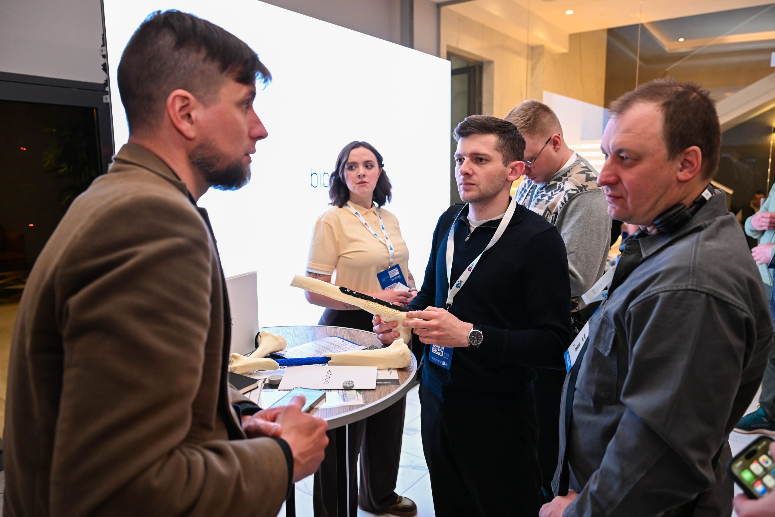
The biodrook team participated in the ASIR conference—one of the key professional events in the field of traumatology and orthopedics, bringing together leading specialists from across Ukraine.

For us, this was not only an opportunity to present our solutions but also a space for meaningful dialogue with doctors who work with complex clinical cases every day.

During the conference, we spoke with practicing surgeons, discussed current challenges in traumatology, and explored the possibilities of integrating custom 3D-printed implants into clinical practice.

Such meetings are of fundamental importance to us—they allow us to gain a deeper understanding of doctors’ needs and develop solutions that are as close as possible to real-world practice.

We are sincerely grateful to the conference organizer, Professor Mykola Ankin, for the high level of organization of the event and for creating a platform for professional exchange.

We valued the opportunity to receive feedback, exchange ideas, and discuss potential areas for future collaboration.

The ASIR conference also served as a platform for establishing new professional contacts. We met a number of specialists

Last week, biodrook welcomed an important guest—Perry Boyle, one of the project’s key investors. This visit provided not only an opportunity to present the current status of production preparations but also to discuss the company’s next stages of development.

The company’s CTO, Mykhailo Pluzhnyk, gave a detailed overview of the progress in setting up the production facility. To date, 90% of the necessary equipment has been purchased, 70% of which is currently being installed. Mykhailo personally coordinates all stages of the process to ensure a smooth and coordinated implementation.

Special attention during the meeting was given to the team. Discussions focused on team growth, adapting to new challenges, and preparing for the full-scale launch of production. For Perry Boyle, this visit was an opportunity to see how the biodrook team works in real time.

This was not just a meeting about technical metrics and plans—it was a moment of mutual trust and support. The visit reminded all participants that biodrook is not just about production capacity, but first and foremost about the people and values on which the company is built. And what lies ahead is just the beginning of a long journey.

In February, the biodrook team held a working meeting with Oleksii Yaremenko, head of the Rehabilitation for Ukraine (Rehab4U) project, and Oleksandr Komarida, head of Objective 1 of this project.

During the meeting, participants discussed potential areas of cooperation in the development of rehabilitation technologies and innovative solutions in regenerative medicine. Particular attention was paid to the potential of modern medical technologies for patient recovery following complex injuries and surgeries

The parties also discussed opportunities to strengthen cooperation between the medical community, technology companies, and research initiatives. Such collaboration can facilitate the faster implementation of innovative solutions in the field of rehabilitation and expand access to modern medical technologies in Ukraine.

The biodrook team values the opportunity for professional dialogue and open exchange of experience. We are convinced that such meetings are an important step toward the development of innovative medicine and the creation of new opportunities for collaboration.

We look forward to further developing our partnership and joint initiatives aimed at supporting and rehabilitating patients in Ukraine.

In February, the biodrook team welcomed a group of leading surgeons and medical specialists to our production facility. The visit marked an important step in strengthening collaboration between clinical practice and biomedical engineering.

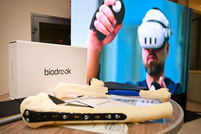
During the meeting, guests were introduced to the full production cycle of patient-specific 3D-printed bone implants: from digital modeling and preparation of biopolymer materials to additive manufacturing and final sterilization. Special attention was paid to the quality control system and process standardization.

The biodrook team demonstrated the capabilities of mobile and stationary production platforms, which enable the rapid fabrication of customized implants. This approach significantly reduces waiting times for patients and increases the predictability of surgical procedures.

The visit served as a platform for open professional dialogue. Surgeons shared practical experience using implants in reconstructive surgeries, and the engineering team received valuable feedback regarding further optimization of the product’s design and clinical integration.

We are convinced that this kind of interdisciplinary collaboration forms the foundation for the development of personalized regenerative medicine in Ukraine and beyond.

The biodrook team has received a Certificate of Conformity and a Project Verification Certificate for our medical device—the anatomically adapted, bioresorbable bone cavity filler ResorBone™ (biodrook).

This means that:

  • our product has been officially confirmed as compliant with the requirements of Ukrainian legislation regarding medical devices;
  • the technical documentation and quality management system have undergone independent verification;
  • the product can be legally manufactured and used in clinical practice in accordance with its intended purpose.

For us, these are not just certificates—they represent an important step in the development of a product created with a focus on patient safety, convenience for doctors, and the trust of the medical community.

At biodrook, we believe that 3D bioprinting is the future of personalized medicine. Our goal is to create custom-made implants that precisely match each patient’s anatomy, are manufactured quickly and affordably, and are eventually replaced by natural bone tissue. This approach reduces the need for additional surgeries and improves the quality of treatment. We are building an innovative bioprinting platform to replace body parts, starting with bone implants—affordably, effectively, and with a patient-centered focus.

Thank you to everyone involved in this process

Moving forward

On December 5, the biodrook team participated in the congress “Amputation and Limb Prosthetics: New Horizons”—an event that holds particular significance today for the entire medical and rehabilitation community in Ukraine.

For us, this congress is not only a platform for exchanging experiences but also a space for shaping a shared vision of the future of prosthetics: technological, accessible, and focused on the real needs of patients. Given the growing number of amputations, the issues of quality, speed, and personalization in prosthetics are becoming critically important.

biodrook sees the future of the industry in the combination of digital technologies, bioengineering, and interdisciplinary collaboration. It is precisely such events that allow us to unite doctors, engineers, prosthetists, and rehabilitation specialists around a common goal—restoring a person’s functionality, dignity, and quality of life.

We are convinced that the development of prosthetics in Ukraine is only possible through dialogue, science, and the implementation of innovations. We thank the organizers and our colleagues for the professional discussion, openness to new solutions, and joint work toward new horizons.

CONTACT US

info@biodrook.com

Start building the future of accessible biotechnology with biodrook today!