
Category: Без рубрики
From Severe Injury to Personalized Reconstruction
The patient sustained a femoral injury in a road traffic accident. She initially received medical care at Kalynivka Central District Hospital. Due to extensive bone damage, she required several stages of surgical treatment. During the first stage, surgeons inserted a temporary bone cement spacer and stabilized the femur using a metal plate and screws. Six weeks later, the spacer was replaced with a patient-specific bioresorbable implant manufactured by the Ukrainian medtech company biodrook. “The femur is one of the largest bones in the human skeleton and plays a crucial role in supporting body weight and enabling movement,” the doctors explained. “Therefore, extensive femoral defects present a significant challenge in reconstructive surgery. Treatment requires not only filling the area of missing bone tissue but also ensuring bone stability, accounting for mechanical loads on the limb, and creating conditions for further regeneration.”What Makes This Approach Innovative?
The uniqueness of this approach lies in the combination of three key elements:- Patient-specific design: The implant was manufactured using 3D printing technology and tailored to the anatomical characteristics of the patient’s bone defect.
- Biodegradation: The implant material is designed to gradually break down within the body.
- Potential for biological regeneration: The treatment aims to achieve the gradual replacement of the implant with the patient’s own bone tissue.
Collaboration Between Surgeons and Medical Technology Specialists
The surgery was performed by a team of surgeons at Vinnytsia City Medical Association No. 2, led by Maksym Verbin and Serhii Kovalenko. An essential part of this treatment approach is close collaboration between medical professionals and technology specialists, from planning the surgical reconstruction to postoperative monitoring and rehabilitation.Patient Recovery and Future Outlook
The patient is currently recovering at home, feels well, and is undergoing rehabilitation. The next stages will involve continued recovery and regular monitoring to assess how the implant gradually degrades and whether it is replaced by the patient’s own bone tissue. The use of personalized 3D-printed implants offers new possibilities for treating complex injuries and supporting bone regeneration. This procedure represents another important step toward integrating advanced reconstructive surgery technologies into medical practice in Vinnytsia.Cranioplasty is a surgical procedure to restore the integrity of the skull bones in the presence of a defect. A defect may result from trauma, tumor removal, an infectious process, or decompressive craniectomy. Restoring the bony coverage of the skull is important not only for protecting the brain but also for restoring normal anatomy and the patient’s appearance.
A patient-specific implant is a medical device created for a particular patient based on their CT data. This approach makes it possible to account for the anatomical characteristics of the defect at the design stage. In this article, we will examine how patient-specific implants differ from standard methods, when their use may be appropriate, what materials they are made from, and how the manufacturing process is carried out.
What is cranioplasty and what does “patient-specific implant” mean?
Cranioplasty is a surgical procedure to restore the integrity of the skull bones in defects of various origins. The aim of the intervention is to provide mechanical protection of the brain, restore normal cerebrospinal fluid circulation, correct the cosmetic defect, and improve the patient’s quality of life.
A patient-specific implant is manufactured according to the anatomy of a particular patient based on computed tomography data. The shape, thickness, and contours of the device correspond to the geometry of the defect and surrounding bony structures before surgery begins.
Unlike standard approaches, in which the surgeon shapes or adapts the material directly during the procedure, a patient-specific implant is created in advance using digital modeling and additive manufacturing technologies (3D printing). This makes it possible to anticipate the reconstruction outcome during the planning stage.
This article focuses on the technology used to create a patient-specific implant: planning, modeling, materials, and the manufacturing process. Surgical techniques for implant placement are beyond the scope of this material.
When is it appropriate to use a patient-specific implant?
A patient-specific implant may be preferred in certain clinical situations. The choice depends on the size and shape of the defect, its location, the condition of the soft tissues, and the clinical goals of reconstruction.
The use of a patient-specific implant may be appropriate in the following cases:
- Large and complex-shaped defects — when the geometry of the defect makes intraoperative modeling technically difficult or time-consuming.
- Cosmetically significant areas — the fronto-orbital region, temporal region, or parietal asymmetry, where accurate restoration of contours is aesthetically important.
- Need for high anatomical accuracy and symmetry — when it is important to restore the natural shape of the skull as precisely as possible.
- Repeat reconstructions — after previous unsuccessful attempts or complications, when a predictable outcome is required.
- Reduction of intraoperative modeling — in situations where shortening the duration of the procedure by using a ready-made device is desirable.
- Interdisciplinary cases — when a neurosurgeon and maxillofacial surgeon work together and a coordinated digital reconstruction plan is required.
Patient-specific implants are most commonly considered for large, irregular, or aesthetically critical defects, as well as for repeat reconstructions.
The final decision regarding the choice of method is made by the operating surgeon after assessing tissue condition, the patient’s overall health, and clinical factors.
How patient-specific implants differ from standard methods
Several approaches are used for cranioplasty. The choice of method depends on the size and location of the defect, the condition of the soft tissues, the clinical objective, and the surgeon’s experience.
Main methods:
- Patient-specific implant — manufactured from the CT data of a specific patient before surgery.
- Titanium mesh — shaped by the surgeon intraoperatively to close the defect.
- Bone cement (PMMA) — shaped directly during surgery.
Comparing these approaches helps to understand the clinical situations in which each may be appropriate.
| Criterion | Patient-specific implant | Titanium mesh | Bone cement (PMMA) |
|---|---|---|---|
| Anatomical accuracy | High; the shape corresponds to the patient’s anatomy based on CT data | Depends on intraoperative modeling by the surgeon | Depends on the shaping technique used during surgery |
| Need for intraoperative modeling | Minimal; the device is ready for implantation | Manual shaping is required during surgery | Material shaping and polymerization are required |
| Duration of surgery | May be shorter because the device is ready-made | Depends on the complexity of the defect and the surgeon’s experience | Depends on polymerization and modeling time |
| Predictability of the outcome | High; the shape can be assessed before surgery | Depends on the surgeon’s technique | Depends on the surgeon’s technique |
| CT/MRI visualization | Depends on the material (PEEK — minimal artifacts, titanium — moderate) | Moderate artifacts | Minimal artifacts |
| Aesthetics in cosmetically significant areas | High due to accurate reproduction of contours | Depends on the shaping technique | Depends on the shaping technique |
| Mechanical stability | High for both materials (PEEK, titanium) | High | Sufficient for most cases |
According to a 2026 systematic review, complication rates for PMMA did not exceed those of titanium implants in the analyzed studies. Some review publications describe fewer imaging artifacts and good clinical outcomes with patient-specific titanium implants.
The choice of cranioplasty method depends on the size and shape of the defect, the condition of the soft tissues, the presence of previous infection or failed reconstruction, and the patient’s cosmetic requirements.
The final decision is made by the physician based on the clinical objective, tissue condition, expected outcome, and available technologies.
Stages of creating a patient-specific implant: from CT images to the finished device
The process of manufacturing a patient-specific implant consists of several sequential stages, each of which is important for ensuring the accuracy of the device and its suitability for the clinical objective.
Main stages:
- Obtaining CT images and preparing DICOM data. The surgeon provides the patient’s computed tomography data in DICOM format (the standard format for medical images). The quality of the source data determines the accuracy of subsequent modeling. Important parameters include slice thickness, resolution, and anatomical coverage.
- Creating a virtual 3D model of the skull and defect. Based on the DICOM data, the engineer creates a digital three-dimensional model of the patient’s skull. The model makes it possible to accurately define the boundaries of the defect, bone thickness, and anatomical landmarks.
- Digital modeling of the implant by the engineer in collaboration with the surgeon; design approval. The engineer designs the implant based on the shape of the defect, skull symmetry, and the surgeon’s clinical requirements. The design is approved by the physician before manufacturing begins, allowing adjustments to be made during the planning stage.
- Manufacturing the device (3D printing or milling) from the selected material. After the design is approved, the implant is manufactured using additive technologies (3D printing, layer-by-layer production) or subtractive methods (milling, machining). The choice of technology depends on the material and device requirements.
- Post-processing, quality control, sterilization, and delivery to the clinic. The finished device undergoes verification of geometric accuracy, mechanical properties, and conformity with the approved design. After sterilization, the implant is delivered to the surgeon together with accompanying documentation.
How biodrook works with the physician during the manufacturing process
biodrook provides technological support at all stages of creating a patient-specific implant.
The collaboration process includes:
- Data exchange — the company provides the physician with requirements for CT data (format, resolution, slice thickness) to ensure the quality of the source information for modeling.
- Co-design with the surgeon — the engineer and surgeon jointly discuss the implant design and agree on its shape, thickness, fixation zones, and other clinically significant parameters.
- Approval protocols — before manufacturing, the surgeon receives the digital implant model for final confirmation.
- Device traceability — each implant is accompanied by documentation recording the stages of manufacturing, quality control, and sterilization.
biodrook reports DSTU EN ISO 13485:2018 certification, indicating the implementation of a quality management system for medical devices. On its product pages, the company also states that implants are tested for geometric accuracy and strength.
This approach allows the surgeon to monitor the process at key stages and receive a device that meets the clinical requirements of a specific patient.
Materials for patient-specific implants: PEEK and titanium
Two materials are most commonly used to manufacture patient-specific cranial implants: PEEK (polyether ether ketone) and medical-grade titanium. Each has properties that may be useful in certain clinical situations.
The choice of material depends on the clinical objective, defect location, imaging requirements, and other factors. There is no universally “best” material for all patients.
PEEK (polyether ether ketone)
PEEK is a biocompatible polymer used in craniofacial reconstruction because of its physical and biological properties.
Key properties:
- Elastic modulus close to that of bone — PEEK has an elastic modulus of approximately 3–4 GPa, which approaches that of cortical bone. This may contribute to more physiological load distribution.
- Radiolucency — PEEK is a radiolucent material and does not produce significant artifacts on CT. This facilitates postoperative monitoring and visualization of brain structures.
- Biocompatibility — the material is generally well tolerated by the body and does not cause pronounced inflammatory reactions in most cases.
- Potential for intraoperative modification — if necessary, a PEEK implant can be further adapted during surgery.
Typical clinical situations:
PEEK may be useful in cases where visualization of brain structures after surgery is important, as well as in patients who require regular CT or MRI monitoring. The material is also considered for reconstruction in cosmetically significant areas because of the ability to reproduce contours precisely.
Medical-grade titanium
Titanium is a metal widely used in orthopedics, traumatology, and craniofacial surgery because of its mechanical and biological properties.
Key properties:
- High strength and stability — titanium has an elastic modulus of approximately 110 GPa, providing high mechanical resistance to direct trauma.
- Proven biocompatibility — titanium has well-documented biocompatibility and a long history of clinical use.
- CT/MRI artifacts — titanium implants may produce moderate imaging artifacts, which should be taken into account when planning postoperative follow-up.
- Thermal conductivity — titanium has higher thermal conductivity than polymers; the clinical significance of this factor is limited for most patients and depends on the individual context.
- Fixation options — titanium implants can be fixed using screws or other fixation systems.
Typical clinical situations:
Titanium may be appropriate in cases where high mechanical stability is required, as well as in patients at risk of repeated trauma. The material is also considered for large defects where long-term structural strength is important.
The choice between PEEK and titanium depends on the clinical objective, defect location, imaging requirements, mechanical requirements, and other factors. The surgeon selects the material based on the patient’s individual characteristics and the goals of reconstruction.
Conclusion
Cranioplasty with a patient-specific implant is a method of restoring the integrity of the skull that makes it possible to account for the anatomy of a specific patient during the device design stage. Patient-specific implants may be appropriate for large or complex defects, cosmetically significant areas, and repeat reconstructions.
The choice between a patient-specific implant, titanium mesh, and bone cement depends on the clinical objective, tissue condition, the surgeon’s experience, and available technologies. Each method has its own advantages and limitations.
The process of creating a patient-specific implant includes obtaining CT data, digital modeling, design approval with the surgeon, manufacturing, and quality control. biodrook provides technological support at all stages and operates according to ISO 13485 quality management system standards.
If you are a physician considering the use of a patient-specific implant for a particular patient, you can contact the biodrook team to discuss the technical feasibility of manufacturing the device and the requirements for the source data.
A gunshot fracture is a bone injury caused by the high-energy impact of a bullet or fragment, accompanied by destruction of bone tissue, wound contamination, and often significant soft tissue defects. Unlike ordinary fractures, such injuries require a specific staged approach: from emergency stabilization to prolonged reconstruction.
This article examines why gunshot fractures require a different treatment strategy, the stages a patient goes through, the fixation and bone defect reconstruction methods used today, as well as the role of modern technologies, including patient-specific 3D implants.
What is a gunshot fracture and why does it require a special approach?
A gunshot fracture occurs when a bullet or fragment penetrates the bone at high velocity. The impact energy damages not only bone tissue but also surrounding soft tissues, blood vessels, and nerves. The wound is always contaminated with particles of clothing, soil, bullet fragments, or shrapnel, creating a high risk of infection.
Key differences from ordinary fractures:
- High-energy injury. The bullet transfers significant kinetic energy to the bone, resulting in a comminuted fracture with a bone tissue defect.
- Wound contamination. A gunshot wound always contains foreign bodies and microorganisms, so the risk of infectious complications is significantly higher.
- Bone and soft tissue defects. Part of the bone tissue may be lost or nonviable. Soft tissues are also damaged, making wound coverage and healing more difficult.
- Combined injuries. Gunshot wounds are often accompanied by damage to blood vessels, nerves, tendons, or joints.
Because of these factors, gunshot fractures of the extremities are managed as open fractures: early intravenous antibiotics, debridement of nonviable tissues and bone fragments, and fracture stabilization are required. There is no universal protocol for all cases: the choice of treatment depends on the fracture location, extent of injury, condition of the soft tissues, and the patient’s overall condition.
Main stages of treatment: from stabilization to reconstruction
Treatment of a gunshot fracture follows a staged approach. The goal of the first stage is to save the patient’s life and limb and to control bleeding and infection. The goals of subsequent stages are to restore bone continuity, cover soft tissue defects, and restore limb function.
The rationale for the staged approach is that performing all tasks simultaneously is often impossible or unsafe. First, the patient’s condition and the fracture are stabilized, then infection is controlled, and only after that is definitive reconstruction undertaken. The individual treatment plan is developed according to the severity of the injury, associated injuries, and the capabilities of the medical facility.
Stage 1. Surgical debridement and temporary stabilization
The first stage of treatment begins with radical surgical debridement of the wound. All nonviable tissue, contamination, foreign bodies, and bone fragments that have lost their blood supply are removed. This is critically important for preventing infection.
After debridement, temporary fracture fixation is performed. External fixation devices are most commonly used, allowing the bone to be stabilized and the length and alignment of the limb to be maintained without requiring large incisions or introducing metal implants into a contaminated wound. External fixation also facilitates access to the wound for repeat debridement.
When indicated, fasciotomy is performed — surgical opening of fascial compartments to reduce soft tissue pressure and prevent compartment syndrome. If the soft tissue defect is extensive, the wound is not closed tightly; instead, early coverage with local or free flaps is planned.
For open fractures, early stabilization is recommended after thorough surgical debridement. In cases of substantial injury or when repeated revisions are necessary, temporary external fixation is preferred, while definitive internal osteosynthesis is performed after safe conditions for soft tissue coverage have been achieved. Definitive soft tissue coverage should be performed as early as possible; if this cannot be done during the initial operation, it should preferably be completed within 72 hours after injury.
Stage 2. Definitive fixation and soft tissue reconstruction
After infection has been controlled and the condition of the soft tissues has stabilized, definitive fracture fixation is performed. If the wound is clean, the soft tissues have been restored, and the fracture is stable, conversion to internal fixation with plates or intramedullary nails may be possible. In some cases, external fixation is maintained until complete union.
The timing and method of soft tissue defect coverage depend on the size and location of the defect. Small defects may heal by secondary intention or be closed with local flaps. Large defects require reconstruction with free flaps using microvascular anastomoses. Early and reliable soft tissue coverage reduces the risk of infection and creates favorable conditions for bone healing.
Reconstruction of the bone defect is often planned as a separate stage once infection has been controlled, the soft tissues have been restored, and the fracture has been stabilized. This may occur several weeks or months after the injury.
Staged treatment pathway for gunshot fractures
- Emergency care: bleeding control, immobilization, antibiotics
- Surgical debridement: removal of nonviable tissue
- Temporary stabilization: external fixation, fasciotomy when indicated
- Infection control: repeat revisions, early soft tissue coverage (preferably within 72 hours)
- Definitive fixation: internal osteosynthesis or prolonged external fixation
- Bone defect reconstruction: selection of method according to the clinical situation
- Rehabilitation: restoration of limb function
Sequence of gunshot fracture treatment stages: from initial debridement to reconstruction
Methods of fixation and bone defect replacement
The choice of fixation method and bone defect reconstruction strategy depends on the fracture location, condition of the soft tissues, infection risks, and the patient’s overall condition. Gunshot fractures often require a separate strategy for the bone defect because part of the bone may have been lost or removed during debridement.
Fracture fixation: external and internal — principles of selection
External fixation devices are appropriate during emergency stabilization when rapid fixation is required without additional trauma to the soft tissues. They allow control of limb length and alignment, provide access to the wound for repeat debridement, and do not require implantation of metal hardware into the contaminated area. Limitations of external fixation include patient discomfort, the risk of pin-site infection, and the need for regular care.
Internal osteosynthesis using plates or intramedullary nails is performed after infection has been controlled and soft tissue coverage has been restored. Internal fixation provides stability, allows earlier weight-bearing, and facilitates rehabilitation. Basic indications for internal osteosynthesis include a clean wound, stable soft tissue condition, and absence of active infection.
Factors that may require a change in strategy include the development of signs of infection, significant bone loss, fixation instability, or deterioration of the soft tissue condition. In such cases, external fixation may be resumed or definitive osteosynthesis may be postponed.
The choice between external and internal fixation depends on the severity of the injury, condition of the soft tissues, and time since injury; the approach is individualized according to the clinical situation.
Bone defect replacement: overview of approaches
When part of the bone is lost or removed, a bone defect develops and requires reconstruction. The main options include:
- Autografting. Transplantation of the patient’s own bone from another site (most commonly the iliac crest). An autograft has the best biocompatibility and osteogenic potential. Limitations include a limited amount of available material, an additional surgical procedure, and pain at the donor site.
- Allograft. Use of donor bone from a tissue bank. An allograft does not require an additional surgical procedure in the patient but has lower osteogenic potential and carries a risk of immunological reactions or infection transmission.
- Induced membrane (Masquelet technique). A two-stage technique: first, a cement spacer is placed in the defect, around which a biological membrane forms; the spacer is then removed and the defect is filled with an autograft. The method is suitable for large defects but requires two operations and prolonged treatment.
- Distraction osteogenesis. Gradual lengthening of the bone using an external fixation device. This method allows reconstruction of substantial defects without bone grafting but requires prolonged use of the device and strict adherence to the distraction protocol.
- Bone substitutes and biomaterials. Synthetic or biological materials that serve as a scaffold for new bone growth. They may be used alone or in combination with an autograft.
Factors considered when selecting a method include the size and location of the defect, infection status, condition of the soft tissues, the patient’s overall condition, resources of the medical facility, and the surgeon’s experience.
Systematic reviews show that autologous bone grafting, the Masquelet technique, and bone transport can provide high union rates in post-traumatic bone defects, but complication profiles and the strength of the evidence vary. High union rates have been reported for autologous bone grafting, although the included studies are heterogeneous. For bone transport, a meta-analysis of large diaphyseal defects showed relatively high union rates but higher rates of complications and repeat procedures compared with grafting techniques. A review of the Masquelet technique for segmental tibial defects indicates that union is usually achieved, often after additional procedures; however, complications are common and direct comparisons with other methods are limited.
| Method | Material/source | When considered | Limitations/risks |
|---|---|---|---|
| Autografting | Patient’s own bone | Small and medium-sized defects, absence of infection | Limited amount of material, donor-site pain, additional surgery |
| Allograft | Donor bone from a tissue bank | Large defects when autograft volume is insufficient | Lower osteogenic potential, risk of immunological reactions, risk of infection |
| Masquelet technique | Induced membrane + autograft | Large segmental defects | Two operations, prolonged treatment, high complication rate |
| Distraction osteogenesis | Patient’s own bone (lengthening) | Substantial defects when grafting is not possible | Prolonged use of the device, high complication rate, need for strict adherence to the regimen |
| Bone substitute materials | Synthetic or biological materials | Small defects, combination with autograft | Limited osteogenic potential, dependence on the specific material |
Methods of bone defect replacement: material source — advantages — limitations/risks
Modern technologies: patient-specific 3D implants in complex cases
For large or complex bone defects, multifragmentary areas, and situations requiring restoration of bone shape and load-bearing function, individualized solutions may be used. Patient-specific 3D implants are created according to the anatomy of a specific patient based on computed tomography data.
The materials used for such implants are most commonly titanium or biocompatible polymers. Titanium has high strength, biocompatibility, and osseointegrative properties. Bioresorbable materials can gradually dissolve in the body, but their use depends on the specific material and clinical objective.
Limitations of patient-specific implants include the need for multidisciplinary planning (surgeon, engineer, manufacturer), cost, manufacturing time, and the need for precise preoperative imaging. A patient-specific implant is not a universal solution for all bone defects but may be useful in complex cases when standard methods are insufficient.
When patient-specific implants may be appropriate
Patient-specific implants are considered in clinical situations where standard solutions are insufficient:
- large bone defects with complex geometry;
- multifragmentary fractures with bone loss;
- defects in anatomically complex regions (pelvis, scapula, proximal extremities);
- the need to restore load-bearing function and anatomical shape;
- inability to use an autograft because of insufficient quantity or the patient’s condition.
Prerequisites for the use of patient-specific implants include infection control, adequate soft tissue coverage, clearly defined reconstruction goals, and the ability to perform preoperative planning based on high-quality CT data. The decision to use a patient-specific implant is made by the surgeon after assessing the clinical situation.
How a patient-specific implant is created: from CT to implantation
The process of creating a patient-specific implant includes several stages:
- Data collection. The surgeon provides the manufacturer with the patient’s computed tomography data.
- Virtual planning. Engineers create a digital 3D model of the bone defect and design an implant that matches the patient’s anatomy. Medical modeling software is used.
- Surgeon approval. The implant model is reviewed and approved by the surgeon. Corrections are made if necessary.
- Manufacturing. The implant is produced using 3D printing on specialized equipment. Selective laser melting or other additive manufacturing technologies are used for titanium implants.
- Sterilization and quality control. The finished implant undergoes sterilization and quality control in accordance with medical standards.
- Intraoperative adaptation. During surgery, the surgeon places the implant and, if necessary, adapts it to the specific clinical situation.
Close collaboration between the manufacturer, surgeon, and medical facility is critically important for a successful outcome. Basic regulatory requirements for medical devices include certification, documentation, instructions for use, and quality control at all stages of production.
The official biodrook website states that patient-specific 3D implants are manufactured from CT/MRI data to fill bone defects after fractures, resections, and reconstructive procedures. The materials undergo preclinical testing in accordance with ISO 10993; the devices are manufactured and supplied sterile. The company reports certification of its quality management system according to DSTU EN ISO 13485:2018 in Ukraine and certification of its products in Ukraine.
From CT scan to placement of a patient-specific 3D implant
- Obtaining the patient’s CT data
- Creating a digital 3D model of the defect
- Designing the patient-specific implant
- Approval of the model by the surgeon
- 3D printing of the implant using specialized equipment
- Quality control and sterilization
- Delivery of the sterile implant to the surgeon
- Intraoperative implantation and adaptation
Workflow for manufacturing a patient-specific implant
Complications and prognosis: what occurs most often and how it is prevented
Gunshot fractures are associated with a high risk of complications. The main risks include:
- Infectious complications. Wound contamination, nonviable tissue, and the presence of foreign bodies create conditions for infection. Deep infection can lead to osteomyelitis — chronic inflammation of the bone that is difficult to treat.
- Nonunion or delayed union. Bone loss, impaired blood supply, infection, and unstable fixation can interfere with fracture healing.
- Loss of soft tissue volume and contractures. Significant soft tissue damage may lead to scar formation, restricted joint movement, and functional impairment.
- Pain syndromes. Chronic pain may result from nerve damage, scar formation, or bone instability.
Factors affecting prognosis include the extent of the initial injury, timeliness and radicality of debridement, stability of fixation, quality of soft tissue coverage, adherence to rehabilitation, the patient’s overall condition, and the presence of comorbidities.
Systematic reviews of gunshot fractures of long bones show that the risk of deep infection and nonunion exists, but the exact frequency varies considerably because of inconsistent definitions of complications and differences in treatment strategies. One review reports generally low infection rates in available case series but emphasizes the lack of standardized definitions and the limited ability to reliably determine the true incidence of deep infection. Complications are most commonly associated with vascular injuries, severe soft tissue damage, bone defects, comminuted or segmental fractures, and high-energy gunshot injuries.
Prevention within the surgical treatment strategy includes staged management, adequate debridement, stable fixation, early functional recovery, and early soft tissue coverage. According to the review, initial intravenous antibiotic therapy is recommended for all affected patients. It is important to understand that thorough debridement remains the standard for high-energy open gunshot injuries.
Rehabilitation: general principles of functional recovery
Rehabilitation after a gunshot fracture is a prolonged process that begins during inpatient treatment. The goals of rehabilitation are pain control, preservation of range of motion, gradual weight-bearing, prevention of contractures and muscle atrophy, and restoration of limb function.
The rehabilitation plan is individualized together with a physical therapist and depends on the fracture location, fixation method, condition of the soft tissues, and the patient’s overall condition. It is important to follow the weight-bearing regimen prescribed by the physician: premature loading may lead to displacement of fragments or fixation failure, while excessive restriction of movement may lead to contractures.
The patient is taught how to use the limb safely, perform exercises to preserve joint mobility, and gradually increase loading. Physical therapy includes passive and active exercises, massage, and physiotherapy procedures when indicated.
Key aspects of communication between the patient, physician, and rehabilitation specialist include regular examinations, radiographic monitoring, adjustment of the weight-bearing regimen according to the rate of bone healing, and discussion of pain and functional limitations. Specific recovery timelines depend on the clinical situation and cannot be determined in advance.
Conclusion
A gunshot fracture is a complex injury that requires a staged approach: from emergency stabilization and infection control to definitive reconstruction of the bone defect and restoration of limb function. The choice of fixation method and bone defect replacement depends on the fracture location, extent of injury, condition of the soft tissues, and the patient’s overall condition.
Modern technologies, including patient-specific 3D implants, expand reconstruction options in complex cases but are not a universal solution. The decision regarding the choice of treatment method is made by the surgeon after careful assessment of the clinical situation.
The Masquelet technique is a two-stage surgical method for treating large segmental bone defects based on the induced membrane phenomenon. The method is used for post-traumatic and post-infectious defects and complex nonunions of long bones when natural bone regeneration is insufficient. The main concept involves creating a biologically active membrane around a temporary spacer, which subsequently provides a favorable environment for bone grafting.
What is the Masquelet technique?
The Masquelet technique is a two-stage surgical method for treating large segmental bone defects. The method was developed for situations in which the amount of lost bone tissue exceeds the capacity for natural regeneration.
The basic principle is as follows: first, a temporary cement spacer is placed in the defect. Over several weeks, an induced membrane forms around it — a biologically active membrane with its own blood vessels and growth factors. During the second stage, the spacer is removed and a bone graft is implanted into the resulting “biological chamber.”
The technique is most commonly used to reconstruct defects of the long bones of the extremities — the femur, tibia, and humerus — after severe trauma, infectious processes, or surgical removal of affected tissue.
The outcome depends on many factors: the size and location of the defect, the condition of the soft tissues, blood supply, presence of infection, stability of bone fixation, and the patient’s overall health. The final decision regarding the choice of method is made by the surgeon after careful assessment of the specific case.
Principle of the method: the induced membrane and its role
The induced membrane is a biologically active membrane that forms around the cement spacer during the first stage of treatment. It performs several important functions that create a favorable microenvironment for subsequent bone formation.
Key functions of the induced membrane:
- Vascularization — the membrane contains numerous blood vessels that provide nourishment to the future bone graft.
- Barrier function — the membrane confines the defect space and prevents bone graft material from dispersing into the surrounding soft tissues.
- Local concentration of growth factors — the membrane produces biologically active substances that stimulate osteogenesis.
In clinical practice, the second stage is usually performed 6–12 weeks after the first, although the optimal interval depends on the location of the defect, the condition of the soft tissues, and the patient’s overall health. There is no single universal protocol suitable for all patients.
Stages of treatment using the Masquelet technique
The Masquelet technique consists of two distinct stages, each with its own clinical objectives. Both stages require surgical stabilization of the bone — internal or external fixation, depending on the location of the defect and the clinical situation.
First stage: debridement of the defect and placement of a cement spacer
During the first stage, thorough debridement is performed — removal of infected, necrotic, or nonviable tissue. This is a critically important step because the quality of debridement directly affects the treatment outcome.
After debridement, a temporary polymethyl methacrylate (PMMA) spacer — bone cement — is placed in the defect. The spacer fills the defect space, maintains bone length, and creates conditions for the formation of the induced membrane.
If infection is present or there is a high risk of infection, antibiotic-loaded PMMA may be used. The decision to incorporate antibiotics into the spacer is made individually. In systematic reviews of infected defects, the use of antibiotic-impregnated cement has been associated with a reduced need for revision procedures and more favorable bone healing outcomes.
At the same stage, stable fixation of the bone segment is ensured and soft tissue coverage of the defect is restored. Without stabilization and adequate soft tissue coverage, the formation of a fully functional membrane is not possible.
Second stage: bone grafting into the induced membrane
The second stage is performed after maturation of the induced membrane — usually 6–12 weeks after the first procedure.
The spacer is carefully removed while preserving the integrity of the induced membrane as much as possible. The membrane remains in place and forms a biological chamber into which bone graft material is implanted.
An autograft — the patient’s own bone, usually harvested from the iliac crest — is most commonly used. Autologous bone is considered the “gold standard” because of its osteogenic properties. In some cases, autologous bone may be combined with allograft (donor bone) or other bone substitute materials, but the choice of graft type is made individually.
After the defect is filled, stable bone fixation is maintained. Further treatment includes monitoring of bone union, rehabilitation, and gradual restoration of limb function.
Key prerequisites for success
Three prerequisites are critically important for achieving a favorable outcome:
- Infection control — radical debridement during the first stage and control of infection before bone grafting.
- Adequate soft tissue coverage — reliable closure of the defect during the first stage.
- Stable fixation — reliable stabilization of the bone segment throughout treatment.
When is the Masquelet technique used: main indications
The Masquelet technique is considered for reconstruction of large segmental defects of long bones in several major clinical scenarios.
Post-traumatic defects — the most common indication for the method. These occur after severe open fractures with loss of bone tissue when the size of the defect exceeds the capacity for natural regeneration.
Post-infectious defects and infected nonunions — situations in which a significant defect remains after surgical debridement of infected bone. The Masquelet technique makes it possible to first control the infection during the first stage and then perform reconstruction.
Defects after tumor resection — cases in which a segmental defect remains after removal of a benign or malignant bone tumor. The suitability of the method is assessed individually depending on the type of tumor and oncological prognosis.
Complex nonunions with bone loss — situations in which previous attempts to treat the nonunion have failed and the amount of lost bone is substantial.
In the literature, a critical-sized bone defect is most commonly defined as a defect longer than 2 cm or loss of more than 50% of the bone circumference. However, there is no single universal threshold, and assessment is always individualized.
The final decision to use the Masquelet technique is made by the surgeon after assessing the condition of the soft tissues, vascular status, infection control, and the ability to provide stable fixation.
Advantages, limitations, and possible complications of the method
The Masquelet technique has several clinically significant advantages, but it is also associated with limitations and risks that must be considered when selecting a reconstruction method.
Advantages
- Possibility of treatment in an infected environment — after thorough debridement, the method allows reconstruction even in post-infectious defects.
- Creation of a biological chamber — the induced membrane provides vascularization and local concentration of growth factors.
- Relative technical accessibility — unlike vascularized grafts, the method does not require microsurgical techniques.
- Compatibility with different fixation methods — both internal and external fixation can be used.
Limitations
- Two operations — the method requires two surgical procedures separated by several weeks.
- Prolonged treatment course — the total time from the beginning of treatment to complete bone union may take several months.
- Need for a sufficient volume of autograft — large defects may require a substantial amount of the patient’s own bone.
- Dependence on fixation stability — without reliable bone stabilization, the outcome is unpredictable.
- Dependence on soft tissue condition — adequate soft tissue coverage is critically important for success.
Possible complications
Systematic reviews show that complication rates with the Masquelet technique vary depending on the location of the defect and the clinical situation. According to different reviews, overall union rates range approximately from 77–96%, depending on location and the presence of infection.
For the lower extremity, some reviews have reported an overall complication rate of approximately 26%, with nonunion (approximately 9%) and deep infection (approximately 8%) being the most common complications.
The most common complications:
- Recurrence of infection — especially in post-infectious defects.
- Nonunion or delayed union — depends on the location and clinical situation.
- Graft resorption — partial resorption of the bone graft material.
- Donor-site problems — pain, infection, or other complications at the site of autologous bone harvesting.
- Fixation-related complications — problems involving metal implants or external fixation devices.
Factors that increase the risk of failure include postoperative infection of the bone graft, age over 65 years, insufficient stabilization, and inadequate debridement during the first stage.
Alternative methods
The Masquelet technique is not the only option for reconstructing large defects. Depending on the clinical situation, the following methods may be considered:
- Ilizarov bone transport — gradual movement of a bone segment to fill the defect.
- Vascularized bone grafts — transplantation of a bone segment together with its blood vessels.
- Combined methods — a combination of different approaches depending on the clinical objective.
The choice of method is always individualized and depends on the size of the defect, its location, the condition of the soft tissues, the presence of infection, and the experience of the surgical team.
The role of 3D printing: patient-specific spacers in the Masquelet technique
Traditionally, cement spacers for the Masquelet technique are shaped manually during surgery. The surgeon molds the PMMA spacer according to the shape and size of the defect. This approach is effective but has potential challenges: variability in shape, difficulty accurately reproducing the anatomy, and inconsistency in spacer volume.
Modern 3D-printing technologies create opportunities for producing patient-specific spacers or molds for shaping PMMA. Based on computed tomography data, an individualized spacer can be designed to more precisely match the anatomy of a specific defect.
Potential advantages of an individualized approach:
- More accurate reproduction of defect anatomy — the spacer can be designed according to the shape and dimensions of the missing bone segment.
- Standardized volume — the required amount of bone graft material for the second stage can be calculated in advance.
- Fixation planning — elements can be incorporated into the spacer design to improve interaction with fixation constructs.
However, it is important to understand the limitations of the evidence: clinical data on the advantages of 3D-printed patient-specific spacers in the Masquelet technique are currently limited. No reliable comparative studies have been identified in the available literature confirming that individualized 3D spacers improve clinical outcomes, reduce complication rates, or increase reconstruction accuracy compared with standard manually shaped PMMA spacers.
The decision to use individualized solutions is made by a multidisciplinary team, taking into account the specific clinical objective, technical capabilities, and safety requirements.
Companies working in the field of medical technology, including biodrook, are developing patient-specific 3D-printed solutions for orthopedics and traumatology. Biodrook manufactures patient-specific bone implants based on CT/MRI data, followed by sterilization and quality control.
What patients should know
If a physician recommends the Masquelet technique for reconstruction of a bone defect, it is useful to understand several key points.
Two-stage treatment — the method involves two operations separated by several weeks. The approximate interval between stages is usually 6–12 weeks, but the specific timing is determined by the surgeon depending on the clinical situation.
Stable bone fixation — throughout treatment, the bone must remain reliably stabilized. This may require the use of metal plates, rods, or external fixation devices. Weight-bearing restrictions on the limb remain in place until complete bone union is achieved.
Rehabilitation and monitoring — after the second stage, regular radiographic monitoring of bone union is necessary. Rehabilitation includes gradual restoration of movement and limb function under the supervision of a physiotherapist.
Infection control — in post-infectious defects, prolonged antibiotic therapy may be required. Adherence to recommendations for surgical wound care is critically important.
Smoking cessation — smoking impairs blood supply and slows bone healing. Smoking cessation significantly increases the chances of a successful outcome.
Individual differences — healing times may vary depending on the size of the defect, its location, the patient’s age, and overall health. In some cases, additional procedures may be required if complications or delayed union occur.
The final decision regarding the choice of treatment method is made by the surgeon after careful assessment of all factors. It is important to discuss expectations, possible risks, and alternative reconstruction options with the physician.
The choice of material for a bone implant affects the outcome of surgery, the ability to monitor bone union, and the long-term stability of the reconstruction. Two of the most commonly used materials — PEEK and titanium — have different properties that determine their applications in orthopedics, spinal surgery, cranioplasty, and maxillofacial reconstruction.
Key points:
- PEEK has an elastic modulus closer to that of bone than titanium, which theoretically reduces the risk of stress shielding.
- Titanium provides natural osseointegration; unmodified PEEK requires surface modification.
- PEEK produces minimal artifacts on CT/MRI; titanium causes pronounced artifacts.
- Titanium has higher fatigue strength for high-load areas.
- The choice depends on the clinical objective, defect location, imaging requirements, and the individual characteristics of the patient.
What are PEEK and titanium in the context of medical implants?
PEEK (polyether ether ketone) is a bioinert high-performance thermoplastic polymer used to manufacture medical implants because of its mechanical properties and chemical stability. PEEK does not actively interact biologically with the tissues of the body.
Medical-grade titanium is a biocompatible metal that has been widely used for implants for decades. Titanium is capable of osseointegration — the formation of direct contact between bone tissue and the implant surface without an intervening connective tissue layer.
Both materials are used in various fields of surgery:
- orthopedics and traumatology — plates, screws, joint prostheses;
- spinal surgery — intervertebral cages, fixation systems;
- cranioplasty — reconstruction of skull defects;
- maxillofacial surgery — reconstruction of facial bones;
- neurosurgery — cranial implants;
- dental implantology — dental implants.
Neither material is universally “better.” The choice depends on the clinical objective, defect location, imaging requirements, expected loads, and the condition of the patient’s tissues.
Key differences: clinically significant properties of PEEK vs titanium
Elastic modulus and the risk of stress shielding
Elastic modulus is a measure of a material’s stiffness. The higher it is, the less the material deforms under load.
PEEK has an elastic modulus of approximately 3–4 GPa, while titanium is approximately 110 GPa; cortical bone is approximately 10–30 GPa. PEEK is closer to bone in terms of stiffness. When an implant is stiffer than bone, it carries a greater proportion of the load. This may lead to stress shielding — a phenomenon in which the bone surrounding the implant receives less mechanical stimulation for remodeling.
Because of its lower stiffness, PEEK is theoretically less prone to stress shielding. However, this does not automatically translate into a clinical advantage in all situations. The impact of stress shielding depends on implant design, anatomical location, type of loading, and the condition of the patient’s bone.
Biocompatibility and osseointegration
Titanium demonstrates a natural ability to osseointegrate. Bone tissue can form direct contact with the surface of a titanium implant, providing stable fixation.
Unmodified PEEK is a bioinert material. Systematic reviews show that it tends to promote the formation of a fibrous capsule (a connective tissue layer) around the implant rather than direct contact with bone. This may limit biological integration.
To improve PEEK osseointegration, surface modifications are used:
- texturing;
- hydroxyapatite coating;
- creation of porous structures;
- combination with titanium components.
Modified PEEK may improve the biological response, but its underlying bioinert nature remains a characteristic of the material. If rapid and reliable osseointegration is critical, titanium has an evidence-based advantage. If bioinertness is acceptable or even desirable, PEEK may be an appropriate choice.
Radiolucency and artifacts on CT/MRI
PEEK produces minimal artifacts on computed tomography (CT) and magnetic resonance imaging (MRI). This allows clear visualization of bone tissue around the implant and assessment of the bone union process after surgery.
Titanium produces pronounced metallic artifacts on CT and MRI. These artifacts can make it more difficult to assess bone condition, detect complications, or plan revision procedures. Titanium is generally considered an MRI-safe material, but local image distortion may limit the diagnostic value of imaging in the area of the implant.
If regular postoperative monitoring of bone union is required or there is a risk of complications requiring imaging, PEEK has an advantage. Most PEEK devices use radiopaque markers to monitor implant position on imaging.
Strength and fatigue resistance
Titanium has high strength and fatigue resistance. It withstands cyclic loading well over long periods, making it a reliable choice for high-load areas.
PEEK has sufficient strength for many applications, but its mechanical properties depend on implant design and loading conditions. In some high-load scenarios, titanium may have an advantage in terms of durability.
In areas exposed to constant high loads (for example, dental implants and certain orthopedic constructs), titanium has a larger evidence base regarding long-term stability.
Machinability and potential for customization
Both materials support modern manufacturing technologies, including 3D printing.
Titanium allows the creation of porous structures that improve osseointegration and load distribution. 3D printing of titanium is widely used for patient-specific implants.
PEEK can also be processed using additive manufacturing technologies. It is easier to machine, providing flexibility in the production of complex geometries.
Both materials make it possible to create customized solutions based on the anatomy of a specific patient.
| Property | PEEK | Titanium | Clinical significance |
|---|---|---|---|
| Elastic modulus | Closer to bone (3–4 GPa) | Significantly higher than bone (110 GPa) | PEEK theoretically has a lower risk of stress shielding; the clinical effect depends on implant design |
| Osseointegration | Requires surface modification | Natural capability | Titanium provides faster and more reliable bone integration |
| Radiolucency | Minimal artifacts | Pronounced artifacts | PEEK allows monitoring of bone union on CT/MRI |
| Strength | Sufficient for most applications | High fatigue resistance | Titanium is more reliable in high-load areas |
| Customization | Supports 3D printing | Supports 3D printing | Both materials allow patient-specific modeling |
Advantages and limitations of each material
PEEK — strengths and limitations
Strengths:
- stiffness closer to that of bone theoretically reduces the risk of stress shielding;
- minimal artifacts allow clear visualization of bone on CT and MRI;
- possibility of customization using 3D printing;
- chemical stability in the biological environment.
Limitations:
- inherent bioinertness requires surface modification to improve osseointegration;
- in its unmodified form, it tends to promote fibrous capsule formation;
- in high-load areas, it may require additional structural support;
- a smaller evidence base compared with titanium for some applications.
Titanium — strengths and limitations
Strengths:
- predictable osseointegration without additional modifications;
- high strength and fatigue resistance;
- extensive clinical experience and evidence base across various fields of surgery;
- ability to create porous structures to improve biological integration;
- reliability in high-load areas.
Limitations:
- pronounced artifacts on CT and MRI make bone visualization more difficult;
- greater stiffness may lead to stress shielding in some scenarios;
- strong osseointegration may make implant removal more difficult during revision procedures;
- its metallic nature may be undesirable for patients with metal sensitivity.
When PEEK is chosen and when titanium is chosen: typical clinical scenarios
When PEEK is more commonly chosen
Spinal surgery — intervertebral cages:
In some clinical practices, PEEK is used for intervertebral cages. Its minimal artifacts make it possible to monitor spinal fusion on follow-up CT scans. Its elastic modulus being closer to that of bone theoretically reduces the risk of stress shielding.
At the same time, systematic reviews and meta-analyses show that 3D-printed titanium cages in the lumbar spine demonstrated better fusion and osseointegration rates compared with PEEK in available comparative studies.
Cranioplasty:
For reconstruction of skull defects, PEEK is often used because it enables the production of patient-specific implants that precisely match the patient’s anatomy. Minimal artifacts allow assessment of bone and soft tissues after surgery.
A meta-analysis of patient-specific craniofacial implants found no statistically significant differences between PEEK and titanium in overall complications, infections, and explantation rates.
Maxillofacial surgery:
In aesthetically sensitive areas with moderate loads, PEEK may be an appropriate choice. It can be shaped according to specific anatomy, and minimal artifacts facilitate postoperative monitoring.
When titanium is more commonly chosen
Dental implantology:
Titanium is the preferred choice in dental implantology and has the largest evidence base for tooth replacement. Its ability to osseointegrate, high strength, and decades of clinical experience make it a reliable option.
High-load orthopedic constructs:
In areas exposed to constant high loads (for example, plates for osteosynthesis of long bones and fixation screws), titanium has an advantage in terms of strength and fatigue resistance.
Situations where rapid osseointegration is critical:
If the clinical objective requires rapid and reliable bone integration with the implant, titanium has an evidence-based advantage. Preclinical data consistently demonstrate better osseointegration of titanium compared with unmodified PEEK.
Hybrid solutions
In some cases, combined approaches are used:
- PEEK implants with titanium coating or titanium components;
- porous PEEK structures with hydroxyapatite coating;
- combination of PEEK and titanium within a single construct.
Such solutions may combine the advantages of both materials, but their use depends on clinical indications and the availability of the required technologies.
These are typical rather than universal scenarios. The final choice of material is an individualized decision made by the surgeon based on clinical data, the evidence base, and the specific characteristics of the patient.
3D printing and patient-specific implants: possibilities for both materials
Additive manufacturing technologies have changed the approach to medical implant production. 3D printing makes it possible to create devices that precisely match the anatomy of a specific patient, which improves load distribution, reduces the risk of complications, and accelerates recovery.
How a patient-specific implant is created
- Obtaining the patient’s medical data — the surgeon provides CT or MRI data in DICOM format.
- Creating a digital 3D model — a three-dimensional model of the anatomical area is generated from the medical data.
- Implant design — engineers develop a construct that takes into account the shape of the defect, mechanical loads, and fixation requirements.
- Approval by the surgeon — the physician reviews the digital model for compliance with clinical requirements and makes adjustments if necessary.
- Manufacturing — the implant is produced using a 3D printer.
- Quality control — the finished device is checked for geometric accuracy, strength, and compliance with specifications.
- Sterilization and packaging — the implant is sterilized and delivered to the surgeon ready for surgery.
Advantages of customization
Patient-specific implants make it possible to:
- accurately reproduce the patient’s anatomy;
- improve load distribution;
- reduce the risk of mismatch between the implant and the defect;
- reduce operating time through preoperative planning;
- create porous structures to improve osseointegration (for titanium).
Possibilities for PEEK and titanium
3D-printed titanium makes it possible to create complex porous structures that mimic cancellous bone. This improves osseointegration and reduces implant stiffness. 3D-printed titanium implants are widely used in spinal surgery, orthopedics, and cranioplasty.
PEEK also supports additive manufacturing technologies. It is easier to machine, providing flexibility in the production of complex geometries. PEEK can be combined with other materials or its surface can be modified to improve the biological response.
Both materials make it possible to create patient-specific solutions, but the choice depends on the clinical objective, indications, and regulatory requirements.
Patient-specific implants from biodrook: a collaborative process with the surgeon
biodrook is a Ukrainian medical technology company that develops and manufactures patient-specific 3D-printed bone implants. The company works as an engineering partner for surgeons, providing a complete cycle from receiving medical data to delivering the finished sterile device.
How biodrook works
- Obtaining medical data — the surgeon provides the patient’s CT or MRI data in DICOM format.
- Creating a digital model — the biodrook team builds a three-dimensional model of the anatomical area and designs the implant.
- Approval by the surgeon — the physician reviews the digital model for compliance with clinical requirements. Adjustments are made if necessary.
- Manufacturing — the implant is produced using biodrook’s own equipment.
- Quality control — the finished device undergoes multilevel verification of geometric accuracy and strength.
- Sterilization and delivery — the implant is sterilized, packaged, and delivered to the surgeon together with the accompanying documentation.
Regulatory compliance
In the European Union, individual patient-specific implants are regulated as custom-made medical devices and must comply with the requirements of Regulation (EU) 2017/745 (MDR). For Class III implantable devices, the conformity assessment procedure under Annex XIII and Annex IX Chapter I applies.
The manufacturer must implement a documented quality management system and prepare technical documentation sufficient for competent authorities to assess the design, manufacturing process, and characteristics of the device. Before the device is placed on the market, the required MDR statement for a custom-made device must be issued.
How the material decision is made: selection factors and the surgeon’s role
The choice between PEEK and titanium is a clinical decision made by the surgeon based on a comprehensive assessment of the situation.
Key selection factors
Defect location:
Different anatomical areas have different implant requirements. For example, in spinal surgery, visualization of bone fusion is important, whereas in dental implantology, osseointegration is critical.
Expected loads:
In high-load areas, titanium may have an advantage in terms of strength and durability. In areas with moderate loads, PEEK may be sufficient.
Imaging requirements:
If regular postoperative monitoring of bone union using CT or MRI is required, the minimal artifacts produced by PEEK are an advantage.
Risk of complications:
If there is a risk of infection, the need for revision surgery, or other complications requiring imaging, PEEK may facilitate diagnosis.
Patient age and comorbid conditions:
Bone condition, the presence of osteoporosis, comorbidities, and the patient’s overall health influence the choice of implant material and design.
Availability of technologies and team experience:
Not all medical facilities have access to 3D printing or experience working with specific materials. The choice also depends on technical capabilities and the expertise of the surgical team.
Principle of shared decision-making
The surgeon explains the available options, their advantages, and their limitations to the patient. The patient has the right to understand why a specific material has been chosen and what trade-offs this involves.
The final decision is made by the surgeon based on clinical data, the evidence base, and the individual characteristics of the patient.
Conclusion:
PEEK and titanium are two different materials with different properties. PEEK has stiffness closer to that of bone and produces minimal imaging artifacts, but requires modification to improve osseointegration. Titanium has a natural ability to osseointegrate, high strength, and extensive clinical experience, but produces pronounced artifacts on CT/MRI.
The choice between them depends on the clinical objective, defect location, imaging requirements, and the individual characteristics of the patient. Neither material is universally “better” — each has its own strengths and limitations.
Modern technologies, including 3D printing, make it possible to create patient-specific implants from both materials, which improves treatment outcomes and reduces the risk of complications. The final decision regarding material selection is made by the surgeon after a comprehensive assessment of the clinical situation.
What is a bone cyst in simple terms?
A bone cyst is a fluid-filled cavity within a bone. It forms in bone tissue, does not contain malignant cells, and does not metastasize. It is a benign lesion that does not transform into a malignant tumor. Bone cysts are most commonly found in children and adolescents aged 5 to 20 years. Typical locations include long bones: the femur, tibia, and humerus. Less commonly, cysts occur in the pelvic bones, spine, or other parts of the skeleton. There are two main types of bone cysts:- Unicameral (simple) bone cyst — the most common type; usually located in the metaphysis (the widened part of the bone near a joint), has a thin wall, and is filled with clear or yellowish fluid.
- Aneurysmal bone cyst — less common; contains blood-filled cavities separated by septa and may increase in size more rapidly.
Why do bone cysts develop and who is at risk?
The exact cause of bone cyst formation has not been definitively established. Several theories have been proposed to explain their development:- Impaired local blood circulation in the bone. Local ischemia or venous obstruction is thought to contribute to cavity formation.
- Abnormalities of bone remodeling. During childhood, bones actively grow and remodel; disruption of this process may contribute to cyst formation.
- Microtrauma. Repeated minor injuries may affect bone structure and create conditions for cavity formation.
- Idiopathic cases. In many patients, no specific cause can be identified.
- Children and adolescents — the main risk group; most cysts are diagnosed between 5 and 20 years of age.
- Boys — unicameral cysts are somewhat more common in boys than in girls.
- Adults — cysts may persist into adulthood or be detected for the first time in adults, but this is less common.
Symptoms of a bone cyst and possible complications
A bone cyst is often asymptomatic. Many patients discover it incidentally — during radiographic examination for another reason or after an injury. Possible symptoms:- Aching pain with weight-bearing on the limb or after physical activity.
- Localized swelling in the area of the cyst if it is located close to the surface.
- Restricted movement or discomfort near a joint if the cyst is large.
- A feeling of weakness in the limb during prolonged physical activity.
- Sudden pain after a minor blow or fall.
- Swelling and deformity of the limb.
- Inability to bear weight on the leg or move the arm.
Pathological fracture — the main danger of a cyst
A pathological fracture is a bone fracture that occurs after minimal trauma or even without trauma due to weakening of the bone structure. How a cyst weakens the bone:- The cavity within the bone reduces the thickness of the cortical layer — the dense outer shell of the bone.
- The larger the cyst and the thinner the wall, the lower the mechanical strength of the bone.
- Under load, the weakened area may fracture.
- A child falls while playing or participating in sports.
- An adolescent sustains a minor blow during everyday activity.
- A patient experiences sudden sharp pain during a normal movement without an obvious injury.
- The size of the cyst — larger cysts carry a greater risk.
- Location — cysts in the lower extremities (femur, tibia) have a higher risk because of constant weight-bearing.
- Thickness of the cortical layer — the thinner the wall, the higher the likelihood of fracture.
- The patient’s activity level — children and adolescents who participate in sports have an increased risk.
How does a physician diagnose a bone cyst?
Diagnosis begins with a consultation with an orthopedic trauma surgeon. The physician takes a medical history and evaluates symptoms, the location of pain, and functional limitations. Diagnostic sequence:- Physical examination and medical history. The physician asks about symptoms, previous injuries, and the characteristics of the pain.
- Radiography — the primary method of initial diagnosis. The cyst appears as a radiolucent cavity with well-defined borders. Radiography makes it possible to assess its size, location, and cortical thickness.
- CT (computed tomography) — provides a more precise assessment of the cyst boundaries and cortical plate, detects occult fractures, and assists with surgical planning.
- MRI (magnetic resonance imaging) — confirms the cystic nature of the lesion, assesses the contents of the cavity, detects septa or fluid levels, and assists with differential diagnosis.
- Radiography does not provide a definitive answer.
- Other lesions need to be excluded.
- Surgical treatment is being planned.
- A pathological fracture or complication is suspected.
Treatment: observation or surgery?
The choice of treatment strategy depends on the patient’s age, the size and location of the cyst, cortical thickness, presence of symptoms, fracture risk, and the patient’s activity level.When observation is sufficient
Active surveillance is used for small asymptomatic cysts with sufficient cortical thickness and a low risk of fracture, particularly if the patient does not participate in contact sports or heavy physical labor. Observation includes regular physician visits, scheduled radiographic or MRI examinations, activity restrictions, and informing the patient about symptoms that require immediate medical attention. A more active treatment approach should be considered if the cyst increases in size, pain appears or worsens, the cortical layer becomes thinner, or a pathological fracture occurs.When surgery is needed: curettage and bone grafting
Surgical treatment is considered for large cysts in weight-bearing areas, symptomatic lesions, a high risk of fracture, pathological fracture with instability, or progression of the cyst during observation. In some cases, a simple cyst may partially or completely heal after a fracture with conservative management; the final decision is made individually by the physician. Curettage is the primary surgical method and involves scraping out the contents and walls of the cyst. The resulting cavity may then be filled with bone grafting material to restore bone strength and support the remodeling process. Bone grafting is the filling of the defect after cyst removal to provide mechanical support and stimulate new bone formation. Materials used for bone grafting:- Autologous bone — the patient’s own bone, usually harvested from the iliac crest; it has high biocompatibility and osteogenic potential but requires an additional incision and may cause pain at the donor site.
- Allogeneic bone — donor bone from a bone bank; it does not require an additional incision but has lower osteogenic activity compared with autologous bone.
- Synthetic biomaterials — artificial materials based on hydroxyapatite, tricalcium phosphate, or composites; they are biocompatible, gradually resorb, and are replaced by the patient’s own bone.
| Management approach | Typical indications | What it includes | Goal | Possible limitations |
|---|---|---|---|---|
| Observation | Small asymptomatic cyst, low risk of fracture | Follow-up visits, scheduled radiography/MRI, activity restrictions | Avoid surgery if the cyst does not progress | Requires patient compliance and regular monitoring; fracture risk remains |
| Surgery (curettage + bone grafting) | Large cyst, symptoms, high risk of fracture, pathological fracture with instability | Removal of cyst contents, filling the defect with bone grafting material | Restore bone strength, reduce fracture risk, relieve symptoms | Surgical intervention, possible complications (infection, cyst recurrence), need for rehabilitation |
Which physician should you consult and what should you do next?
A bone cyst is treated by an orthopedic trauma surgeon. For children, consultation with a pediatric orthopedic trauma surgeon experienced in pediatric bone disorders is recommended. Seek immediate medical attention in the following situations:- Sudden severe pain in a limb after minor trauma or without an obvious cause.
- Limb deformity, swelling, inability to bear weight on the leg or move the arm.
- Suspected pathological fracture.
- Previous radiographs or CT/MRI reports, if available.
- A list of medications the patient is taking.
- A brief history of symptoms — when the pain started, whether there was an injury, and how the symptoms have changed.
Conclusion
A bone cyst is a benign lesion most commonly found in children and adolescents. The main danger is weakening of the bone and the risk of pathological fracture. The choice between observation and surgical treatment depends on the size of the cyst, its location, symptoms, cortical thickness, and the patient’s activity level. For small asymptomatic cysts with a low risk of fracture, active surveillance may be sufficient. For large cysts, significant fracture risk, or the presence of symptoms, surgical intervention — curettage followed by bone grafting — may be considered. The final decision regarding treatment strategy is made by an orthopedic trauma surgeon after a complete examination and assessment of the clinical situation. If you or your child has been diagnosed with a bone cyst, consult an appropriate specialist for a detailed evaluation and selection of the optimal treatment approach.What is “artificial bone” in orthopedics and surgery?
Artificial bone is not a single product but a group of bone substitute materials with different properties and forms. They are used for two main purposes:- Defect replacement and structural support. The material fills a cavity or restores lost bone volume, providing mechanical support.
- Stimulation of the patient’s own bone growth. Some materials create a scaffold along which new bone can grow. This process is called osteoconduction — the ability of a material to serve as a structure along which new bone tissue forms.
When is bone tissue reconstruction needed: main indications
Bone tissue reconstruction may be indicated in various clinical situations:- Post-traumatic defects. After severe fractures, especially open fractures or those involving loss of bone fragments.
- Consequences of tumor or cyst removal. The cavity left after surgical removal of pathologically altered tissue requires filling.
- Congenital or postoperative defects. Bone deformities that require correction and restoration of shape.
- Reconstruction in maxillofacial surgery. Restoration of jaw bone tissue, including preparation for placement of dental implants.
Artificial bone materials: bioresorbable and bioinert
Bone substitutes are classified according to their behavior in the body: materials that are resorbed and materials that remain as long-term structural support. Any material must meet two basic requirements:- Biocompatibility. The material must not cause a toxic or immune reaction.
- Suitability for the clinical task. The shape, volume, and mechanical properties of the material must be appropriate for the specific defect and load.
Bioresorbable materials: a scaffold for the growth of the patient’s own bone
Bioresorbable materials gradually resorb in the body and are replaced by the patient’s own bone tissue. They create a temporary osteoconductive scaffold — a structure along which new bone can grow. Indications:- small and medium-sized defects;
- areas with adequate blood supply;
- situations where natural bone repair and remodeling are expected.
Bioinert materials: structural support without resorption
Bioinert materials are not resorbed in the body. They retain a stable shape and volume, providing long-term mechanical support. Depending on the material, direct bone contact — osseointegration (a direct structural connection between bone and the implant surface) — may occur, but replacement by the patient’s own bone does not take place. Indications:- large defects requiring permanent support;
- areas exposed to high mechanical loads;
- situations where restoration of shape is more important than biological integration.
Forms of bone substitutes: from granules to patient-specific implants
Bone substitutes are available in different forms, each suited to specific clinical tasks. Common forms:- Granules or chips. Used to fill irregularly shaped cavities. The granules can be tightly packed to fill the defect.
- Pastes or gels. Convenient for injection using a syringe. Suitable for small defects or for filling complex anatomical areas.
- Blocks or plates. Used to restore bone contour and volume. A block can be shaped during surgery or manufactured in advance.
Patient-specific 3D implants: when they are appropriate and how they are created
Patient-specific 3D implants are personalized medical devices developed based on computed tomography or MRI data from a specific patient. They are designed to match the anatomy of the defect, helping restore its shape according to preoperative planning and improving the fit of the implant to the defect. Indications:- complex or large bone defects;
- the need for precise restoration of anatomical shape;
- reconstruction after tumor removal or trauma involving loss of a substantial amount of bone;
- cases where standard material forms do not meet the clinical requirements.
Stages of planning and manufacturing
The process of creating a patient-specific implant includes several sequential stages:- Obtaining CT or MRI data. The physician provides medical images for analysis.
- Creating a 3D model of the defect. A digital model of the anatomical area is created based on imaging data.
- Virtual planning. The surgeon and engineer jointly agree on the shape, dimensions, and position of the implant.
- Manufacturing or printing. The implant is produced using specialized equipment and additive manufacturing technologies.
- Sterilization and preparation. The finished device undergoes the necessary procedures to ensure sterility.
- Surgical placement. The implant is provided to the surgeon for the procedure.
- Follow-up. Postoperative imaging is performed to assess the position of the implant.
Example of a workflow with patient-specific implants
biodrook integrates the patient’s CT data, coordinates the implant design with the surgeon, and manufactures the device according to the patient’s individual anatomy. If necessary, surgical guides can be prepared for precise positioning of the implant during surgery. The process includes a complete technological cycle: from digital modeling to the finished medical device. Important: biodrook does not make clinical decisions. Indications for the use of a patient-specific implant, the choice of material, and the treatment strategy are determined by the physician after examining the patient.Advantages and limitations: artificial bone vs the patient’s own bone (autograft)
An autograft — the patient’s own bone taken from another area of the body — is traditionally considered the reference option for bone grafting. ISASS recommendations emphasize that the patient’s own bone is the reference standard in spinal surgery, while substitutes are used as an alternative or adjunct, taking into account their mechanisms of action and limitations. Autografts have limitations that make artificial materials a reasonable alternative in a number of clinical scenarios.| Criterion | Artificial bone materials | Autograft |
|---|---|---|
| Additional surgical site | Not required | Required (donor site) |
| Volume limitations | Depends on the manufacturer | Limited available volume |
| Biological potential | Depends on the material; osteoconduction is possible | High; contains living cells |
| Predictability of shape | High; a precise shape can be manufactured | Limited; depends on the donor site |
| Risk of donor-site complications | Absent | Pain, infection, impaired function |
| Risk of surgical-site infection | Depends on many factors; there is no risk of transmission of donor infections | There is no risk of infection transmission from a donor; surgical infections are possible |
| Integration time | Depends on the material and clinical situation | May be faster; depends on the clinical situation |
- Autograft has high biological potential but requires additional surgical trauma and is limited in volume.
- Artificial materials make it possible to avoid a donor site, provide a predictable shape, and can be manufactured in the required volume.
- For large or complex defects, artificial materials, particularly patient-specific implants, may be one of the options depending on the clinical situation and indications.
Possible risks and complications
The use of bone substitutes, like any surgical intervention, is associated with risks:- surgical-site infection;
- incomplete defect replacement or nonunion;
- need for revision surgery;
- rare individual reaction to the material.
How does the physician choose the optimal material and method?
The choice of bone tissue reconstruction method is an individualized decision made by the physician after a comprehensive assessment of the clinical situation. Key criteria:- Size and location of the defect. Small defects may be filled with granules or pastes; large defects require blocks or patient-specific implants.
- Load on the area. Areas subjected to high mechanical loads require materials with appropriate strength.
- Age and comorbid conditions. The patient’s overall health, presence of chronic diseases, and blood supply influence the choice of method.
- Healing time. Some materials integrate faster, while others require a longer recovery period.
- Goals of the procedure. Restoration of function, aesthetics, or both.
- Availability of tissues and technologies. The possibility of harvesting an autograft and the availability of the necessary equipment and materials.
September at biodrook: Healthcare, Technology, Partnerships and New Connections
September was a month of active engagement for biodrook. Throughout the month, our team joined healthcare and technology events, met with clinicians, investors and business leaders, strengthened international connections, and continued building a stronger dialogue around patient-specific medical technologies.1. Connecting with Ukraine’s business ecosystem
We started the month at Welcome Day by the Union of Ukrainian Entrepreneurs (SUP). The event brought together members of Ukraine’s business community and created an opportunity to discuss potential partnerships and new areas for collaboration. For biodrook, connections beyond the medical sector are an important part of growth. Building a medical technology company also means being part of a wider ecosystem of entrepreneurs, technology companies, investors and partners.
2. Healthcare reconstruction and the future of Ukrainian MedTech
At Ukraine ReHealth 2026, biodrook joined an international dialogue between government representatives, investors, healthcare professionals and businesses focused on the recovery and future development of Ukraine’s healthcare system. For our team, the event was an opportunity to better understand the priorities shaping healthcare reconstruction and the potential role of Ukrainian MedTech in this process.
3. Investment and the future of Ukrainian technology
On 9 September, we joined FutureTech Meetup & InvestDay by Diia.City United, focused on investment in Ukrainian technology and the future of innovation. Meeting investors and representatives of the Ukrainian technology ecosystem allowed us to explore different approaches to partnerships, investment and international growth. These connections are particularly important as biodrook continues working toward bringing Ukrainian patient-specific medical technologies to broader markets.
4. Digital medicine meets patient-specific technology
Digital Medicine Forum 2026 in Kyiv brought together more than 250 participants and 30+ speakers from healthcare, MedTech, IT, eHealth and the public sector. The discussions around AI, telemedicine, digital radiology and medical data were especially relevant to biodrook. Our own workflow starts with a patient’s CT or MRI data and transforms digital medical information into a sterile, patient-specific medical device. As healthcare becomes increasingly digital, new opportunities emerge for integrating personalized technologies into everyday clinical practice.
5. Staying close to the clinical community
On 18 September, biodrook attended the scientific and practical conference of the Ukrainian Association of Sports Medicine in Kyiv. Sports injuries, arthroscopy, reconstructive orthopedics and patient rehabilitation were among the key topics. For our team, participating in specialized medical events is an opportunity to remain close to clinicians and better understand where personalized technologies and patient-specific solutions may support complex orthopedic and trauma care. September 2026 Update
6. Building a better customer experience
Technology alone does not define the experience of working with biodrook. On 22 September, our team joined the 21’ Customer Experience Conference online to explore personalization, human-centered service, employee experience, AI-first approaches and digital transformation. Our work requires close interaction with surgeons and medical teams throughout the entire process, from clinical data and implant design to production, surgery and post-market follow-up. As biodrook grows, making this experience more consistent and effective remains an important part of our development. September 2026 Update
7.Representing Ukrainian MedTech internationally
On 25 September, biodrook attended the Business, Innovation, Science and Trade reception organized by the British Embassy in Ukraine as part of IT Arena in Lviv. The event brought together representatives of business, science, innovation and international organizations. biodrook participated as a representative of Ukrainian MedTech, contributing to a broader dialogue around technology, science, innovation and international cooperation. September 2026 Update
8. IT Arena 2026: looking beyond MedTech
From 25 to 27 September, our team attended IT Arena 2026 in Lviv, one of Ukraine’s major technology events. AI, product development, investment, emerging technologies and global scaling were among the topics particularly relevant to biodrook. One of our key takeaways from IT Arena was simple: strong technology is only the beginning. Creating sustainable impact also requires the right product strategy, partnerships, investment and readiness to scale. We left IT Arena with new connections, perspectives and ideas to bring into the further development of biodrook.
9. Exploring new opportunities in dentistry
On 26 September, biodrook attended Ukrainian Dental Forum 2026 in Kyiv, which brought together dentists, clinic owners and leaders of the dental industry. The forum provided insights into treatment quality, patient experience, clinic management and the adoption of new technologies in dentistry. We were also glad to meet and exchange ideas with Yaroslav Kazio and Ihor Kazio. Direct dialogue with clinicians helps us better understand real clinical needs and identify where our expertise in 3D printing and patient-specific medical devices can create value in new areas of medicine.
10. A new space for patient voices
September also brought an important milestone for biodrook: the launch of our dedicated Patient Reviews page. The page brings together video testimonials and experiences from patients whose treatment involved biodrook patient-specific solutions. It complements the clinical cases, physician feedback and follow-up information we collect by adding another important perspective: the experience of the patient. As we document more cases and long-term follow-ups, this page will continue to grow. September 2026 Update Read patient reviews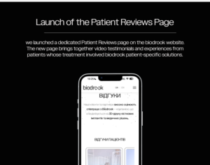
What is a false joint in simple terms?
A false joint is the result of impaired fracture healing. Normally, bone tissue is capable of regeneration: after a fracture, a bone callus forms between the fragments and gradually develops into mature bone. If this process stops or does not proceed properly, a gap remains between the fragments, and abnormal mobility develops at the fracture site — the so-called false joint. Pseudarthrosis is diagnosed when a fracture fails to unite within the expected period of consolidation and the absence of healing progression is confirmed radiologically. This is not merely delayed union — it is a condition in which the biological conditions required for consolidation have been lost, and the likelihood of spontaneous union is low. Surgical intervention is generally required. Potential consequences of pseudarthrosis:- chronic pain at the fracture site;
- loss of weight-bearing function or limb strength;
- deformity and malalignment of the limb;
- risk of disability and the need for prolonged treatment;
- possibility of infectious complications.
How to recognize and confirm the diagnosis: symptoms and examinations
Pseudarthrosis is suspected when symptoms do not resolve within the clinically expected timeframe after a fracture and the patient’s condition does not improve. Main symptoms:- persistent pain at the fracture site that does not decrease over time;
- abnormal mobility at the site where the bone should have united;
- swelling, deformity, or a change in the shape of the limb;
- weakness, loss of function, inability to fully bear weight on the limb;
- absence of clinical and radiological progression of healing.
Why the bone does not heal: main causes and risk factors
Successful fracture healing requires four conditions: mechanical stability of the fragments, adequate blood supply, viable cells, and absence of infection. If even one of these conditions is impaired, the risk of pseudarthrosis increases. Current reviews and AO Foundation materials emphasize that the risk of nonunion depends on fracture severity and the quality of restored stability, so these factors should be assessed together rather than in isolation. Causes and risk factors can be divided into two groups: those related to the injury and treatment, and those related to the patient’s overall health.Factors related to injury and treatment
Mechanical instability is one of the most common causes of pseudarthrosis. If the bone fragments are not adequately stabilized, continuous movement at the fracture site prevents bone callus formation. This may occur due to insufficient or inappropriate fixation, breakage or migration of fixation hardware, or premature weight-bearing on the limb. Large bone defects or loss of a bone segment after injury create a gap that the body cannot fill on its own. The larger the defect, the greater the need for biological stimulation of bone union. Infection (osteomyelitis) disrupts the normal healing process. Infected tissues cannot provide the conditions required for consolidation, so the infection site must first be debrided before stability can be restored. An open fracture increases the risk of pseudarthrosis because of soft tissue damage, impaired blood supply, and possible infection. More severe fracture morphology (comminuted, oblique, spiral) makes stable fixation more difficult to achieve and increases the risk of nonunion.Factors related to the patient’s overall health
Smoking is a proven risk factor for pseudarthrosis. Nicotine impairs blood supply to bone tissue and reduces the activity of osteoblasts — the cells responsible for forming new bone. Diabetes mellitus impairs microcirculation and slows regenerative processes. Patients with poorly controlled diabetes have a higher risk of nonunion. Obesity is associated with an increased risk of pseudarthrosis, although the mechanism behind this association is not fully understood. It may be related to chronic inflammation, metabolic disturbances, and mechanical loading of the fracture site. Impaired blood supply at the fracture site is a critical factor. If the bone does not receive sufficient oxygen and nutrients, the healing process stops. A 2022 meta-analysis showed statistically significant associations between nonunion and smoking, diabetes mellitus, obesity, open fractures, and greater injury severity according to classification systems.Types of pseudarthrosis and why they matter for treatment
Pseudarthroses are classified according to radiological and biological features. The most common classification divides them into two main types: hypertrophic and atrophic. Understanding the type of pseudarthrosis helps determine the appropriate treatment strategy. Hypertrophic pseudarthrosis is characterized by excessive bone formation. Radiographs show pronounced callus formation around the fracture site, but the fragments have not united because of insufficient stability. The biological healing potential is preserved, but the mechanical conditions do not allow the consolidation process to be completed. Treatment goal: provide stable fixation. Osteosynthesis alone is often sufficient without additional bone grafting because the biological capacity for union remains intact. Atrophic pseudarthrosis is characterized by absent or minimal callus formation. The ends of the fragments are sclerotic, blood supply is impaired, and the biological healing potential is reduced or lost. It is often accompanied by a bone defect. Treatment goal: not only stabilization, but also biological stimulation of union. This may include bone grafting, use of osteoconductive or osteoinductive materials, and restoration of blood supply.| Characteristic | Hypertrophic pseudarthrosis | Atrophic pseudarthrosis |
|---|---|---|
| Radiological features | Pronounced callus, “elephant foot” appearance | Absence of callus, sclerosis of the fragment ends |
| Biological potential | Preserved | Reduced or lost |
| Main problem | Fixation instability | Poor blood supply, bone tissue deficiency |
| Treatment focus | Stabilization | Stabilization + biological stimulation |
Modern treatment approaches: how bone union is restored
Surgical treatment is the primary approach for established pseudarthrosis. Success depends on infection control (if present), achieving stability, and restoring a favorable biological environment. Conservative methods (immobilization, physiotherapy) have limited effectiveness and do not eliminate the main causes of nonunion. The general principle of pseudarthrosis treatment is based on three components:- Elimination of infection (if present). Infected pseudarthrosis requires a staged approach: first, debridement of the infection site, removal of nonviable tissue, microbiological confirmation, systemic and, if necessary, local antibiotic therapy, followed by definitive stabilization.
- Restoration of mechanical stability. Bone union is impossible without stable fixation of the fragments.
- Provision of biological conditions for union. This may include bone grafting, the use of osteoconductive materials, and restoration of blood supply.
Surgical treatment: osteosynthesis and bone grafting
Osteosynthesis is the surgical fixation of bone fragments using metal implants. The choice of fixation method depends on the location of the pseudarthrosis, the type of fracture, and the condition of the bone:- Plates and screws provide rigid fixation, particularly for metaphyseal fractures.
- Intramedullary fixation (nails) is commonly used for diaphyseal fractures of long bones.
- External fixation may be used for infected pseudarthrosis or complex defects.
- Autografts (the patient’s own bone, usually harvested from the iliac crest) — the “gold standard” because they contain living cells and growth factors. The disadvantage is the need for an additional surgical approach and the limited amount of available material.
- Allografts (donor bone) — do not contain living cells but provide osteoconduction (a scaffold for new bone growth). The risk of an immune reaction is minimal after processing.
- Synthetic osteoconductive materials (calcium phosphate ceramics, bioactive glass) — provide a scaffold for bone growth and are gradually resorbed. In a meta-analysis of nonunion treatment, synthetic substitutes demonstrated an overall union rate of approximately 90%, although outcomes vary depending on the material and study design.
- Infected sites require debridement first.
- Large segmental defects may exceed the capabilities of standard bone grafting.
- Compromised blood supply or severe comorbidities reduce treatment effectiveness.
- Complex cases may require a staged or combined approach.
Patient-specific 3D-printed implants for bone defects
For significant bone defects, standard methods may be insufficient. In such cases, patient-specific 3D-printed implants may be considered. Indications for patient-specific solutions:- segmental defects or loss of bone tissue;
- complex anatomy where standard implants do not match the geometry of the defect;
- the need to restore limb length, alignment, or weight-bearing function;
- combining fixation with bone grafting or soft tissue reconstruction.
- A patient-specific implant must be designed with biomechanical loads and the clinical objective in mind.
- The implant material must meet regulatory requirements and be biocompatible with biological tissues.
- Active infection must be excluded before implantation.
- Patient-specific solutions do not eliminate the need for stable fixation and biological stimulation of bone union.
Recovery and prognosis: what to expect after surgery
After surgical treatment of pseudarthrosis, the patient needs time for bone consolidation and restoration of limb function. Stages of recovery:- Early postoperative period: pain management, wound care, and monitoring of soft tissue condition.
- Immobilization or weight-bearing restrictions as needed, depending on the location and type of fixation.
- Gradual mobilization under medical supervision to prevent contractures and maintain circulation.
- Follow-up radiographs to assess the progression of bone union.
- delayed union or recurrent nonunion;
- infectious complications;
- breakage or migration of fixation hardware;
- need for repeat surgical procedures.
When to consult an orthopedic trauma surgeon
Timely consultation with an orthopedic trauma surgeon is critically important for successful treatment of pseudarthrosis. Consult a physician if:- pain and impaired limb function persist after a fracture beyond the expected healing period;
- there is no progress on follow-up radiographs;
- abnormal mobility develops at the fracture site;
- limb deformity develops;
- there are signs of infection: redness, swelling, wound discharge, fever.
Key points at a glance
- Delayed union — bone healing is progressing more slowly than expected, but the process is continuing. NICE benchmark for long bones: no radiographic signs of union after 3 months.
- Nonunion (pseudarthrosis) — the healing process has stopped. NICE benchmark and FDA labeling: at least 9 months have elapsed, with no visible progression of healing during the last 3 months.
- The strongest evidence-based risk factor is infection (the odds of nonunion are 7 times higher). This is followed by open fracture, smoking, diabetes mellitus, and obesity.
- Symptoms that should raise concern: pain that does not decrease; pain with weight-bearing; abnormal mobility; failure of fixation hardware.
- The diagnosis is made by a physician based on examination and serial radiographs or CT — not on timelines found on the internet.
- For established pseudarthrosis, treatment is usually surgical: revision osteosynthesis, infection debridement, and bone grafting. In complex cases, patient-specific 3D-printed implants may be considered — an approach with a currently limited evidence base.
What are delayed union and nonunion (pseudarthrosis)?
When a bone breaks, the body initiates fracture healing. Normally, the process occurs in stages: a bone callus forms, the fragments gradually unite, and after a certain period the fracture heals. In some cases, the process slows down or stops. Delayed union is a condition in which healing occurs more slowly than expected for the specific bone and type of fracture, but the process is still ongoing. The bone continues to form callus, although with a delay. Fracture nonunion (nonunion, pseudarthrosis) is a condition in which healing has stopped and spontaneous union is unlikely without intervention. A false joint may form at the fracture site, with persistent abnormal mobility between the bone fragments.How does delayed union differ from nonunion?
| Feature | Delayed union | Nonunion (pseudarthrosis) |
|---|---|---|
| Status of the process | Ongoing, but slow | Stopped |
| Approximate timeframe (long bones) | No signs of union after ~3 months | ≥9 months since injury, with no progress during the last 3 months |
| Serial radiographs | Callus formation is delayed | No change between radiographs |
| Prognosis without intervention | Union is possible | Spontaneous union is unlikely |
| Main management approach | Correction of risk factors, optimization of fixation and loading; observation | Usually surgical treatment |
Why does a fracture fail to heal?
The causes are divided into two groups: systemic factors, related to the overall condition of the body, and local factors, directly related to the fracture and its treatment. A combination of several factors is often involved, and the more factors that are present simultaneously, the higher the risk.How strong is each risk factor?
A systematic review and meta-analysis of 30 studies (Jensen et al., EFORT Open Reviews, 2022) assessed risk factors for nonunion after surgically treated diaphyseal fractures:| Risk factor | Odds ratio (OR) | 95% CI |
|---|---|---|
| Infection | 7,0 | 3,2–15,0 |
| Open fracture | 2,6 | 1,8–3,9 |
| Severity according to the AO classification | 2,4 | 1,5–3,7 |
| Severity according to the Gustilo classification | 2,2 | 1,4–3,7 |
| Smoking | 1,7 | 1,2–2,4 |
| Diabetes mellitus | 1,6 | 1,3–2,0 |
| Obesity | 1,5 | 1,1–1,9 |
Systemic factors
Smoking and nicotine exposure. A separate systematic review and meta-analysis (122 studies, 417 767 patients; Xu et al., EClinicalMedicine, 2021) showed that smokers had a higher rate of nonunion — OR 2,50 (95% CI 1,73–3,61). Nicotine impairs tissue blood supply and reduces oxygenation. An important practical detail: smoking cessation at least 4 weeks before surgery was associated with a lower rate of postoperative wound infection (OR 0,37). Diabetes mellitus. Impaired microcirculation and altered collagen synthesis reduce the body’s ability to restore bone structure. Glycemic control is an important component of preparation for treatment. Infection at the fracture site. According to available data, this is the strongest risk factor for nonunion. The infectious process damages tissues and sustains chronic inflammation, which interferes with bone healing. Obesity. It places additional mechanical stress on the bone and is often accompanied by metabolic disturbances that affect healing. Vitamin D, calcium, and protein deficiency. These nutrients are necessary for bone tissue formation. Correction should be prescribed by a physician based on laboratory findings, rather than “just in case.” Osteoporosis. Reduced bone mineral density may make the formation of a strong callus more difficult, although direct evidence of this association remains limited. Age. In older patients, regenerative processes occur more slowly, while comorbidities may further complicate healing. Use of certain medications. The EAST/OTA clinical guideline on pain management after orthopedic trauma states that the evidence regarding NSAIDs and nonunion is conflicting. Intravenous ketorolac during the acute period improved pain control and reduced opioid requirements without increasing the rate of nonunion. At the same time, prolonged use of high-dose NSAIDs should be discussed with a physician.Local factors
Type and severity of injury. Open fractures, comminuted fractures, fractures with major displacement, or fractures with bone loss carry a higher risk. Soft tissue and vascular damage complicates healing. Impaired blood supply to the fragments. Loss of normal blood supply to one or both bone fragments slows or stops callus formation. Insufficient or unstable fixation. Mobility of the fragments interferes with the formation of a strong callus. Stable fixation is one of the main prerequisites for successful union. Soft tissue interposition. Entrapment of muscles, tendons, or other tissues between bone fragments mechanically prevents union. Large bone defect. A substantial defect may exceed the body’s biological capacity for spontaneous repair.Which symptoms should raise concern?
- Persistent pain at the fracture site — pain that does not decrease over time or becomes more severe.
- Pain with weight-bearing — attempting to bear weight on the limb or perform movement causes pain at the fracture site.
- Persistent swelling or deformity — swelling that does not resolve or a visible change in the shape of the limb.
- Abnormal mobility — a feeling of instability where the bone should already have healed.
- Grinding or clicking during movement.
- Lack of functional improvement — the ability to move normally or bear weight on the limb does not recover within the expected timeframe.
- Breakage or loosening of fixation hardware. If a metal implant breaks, this may indicate that the bone has not united and the entire load is being carried by the hardware.
How does a physician diagnose impaired fracture healing?
Clinical examination. The physician evaluates symptoms, limb function, pain, swelling, deformity, and abnormal mobility. It is important to assess how the symptoms have changed over time. Serial radiography. Radiographs are obtained at specific intervals. The physician assesses whether signs of callus formation are appearing and whether the fracture line is becoming less visible. If the appearance remains unchanged for several months, this indicates nonunion. Computed tomography (CT). Provides more detailed information about callus quality, persistence of the fracture line, the condition of the fixation, and bone defects. It is particularly useful for surgical planning. Evaluation for infection. If an infectious process is suspected, laboratory tests, tissue cultures, or other studies are performed to identify the causative pathogen.Hypertrophic or atrophic: why does it matter?
| Hypertrophic nonunion | Atrophic nonunion | |
|---|---|---|
| Radiographic appearance | Excessive bone formation around the fracture line, but without union | No bone formation; the ends of the fragments are “smoothed” |
| Primary cause | Mechanical: excessive mobility, unstable fixation | Biological: impaired blood supply, cellular deficiency |
| What this means | The biology is functioning, but the mechanics are interfering | The mechanics may be adequate, but the biology is not functioning |
| Main treatment approach | Improving fixation stability | Biological stimulation: bone grafting, restoration of blood supply |
What treatment options are available?
The choice depends on the type of fracture, type of nonunion, fixation stability, presence of infection, size of the bone defect, and the patient’s overall condition.Can nonunion be treated without surgery?
Conservative treatment may be appropriate for delayed union when there are signs that the healing process is continuing. In such cases, the physician may recommend: Correction of modifiable risk factors. Smoking cessation, glycemic control in diabetes, correction of laboratory-confirmed vitamin D and calcium deficiency, and optimization of nutrition. Optimization of immobilization and weight-bearing. The physician may adjust the weight-bearing regimen or method of fixation. Biostimulatory methods. In the NICE MTG12 guidance, low-intensity pulsed ultrasound therapy (LIPUS, EXOGEN system) is considered an option for nonunion and delayed healing of long bones when appropriate indications are present. It is prescribed individually by a physician; it does not replace surgical intervention in established pseudarthrosis. Limitations of the approach. In established pseudarthrosis, when the healing process has stopped, conservative methods are usually insufficient.Surgical approaches to nonunion
Surgical treatment is based on two principles: stable fixation and biological stimulation of union. If infection is present, controlling it is a mandatory prerequisite. Revision osteosynthesis. If insufficient stability is the cause, revision surgery is performed: replacement or reinforcement of fixation devices, or a change in the type of fixation (for example, switching from a plate to an intramedullary nail or vice versa). Infection management. Removal of infected tissue, debridement of the fracture site, and, if necessary, antibacterial therapy. The infection must be controlled before or during reconstructive surgery. Bone grafting. Addition of bone tissue to the fracture site to stimulate union.Autograft, allograft, or synthetic material?
| Autograft | Allograft | Synthetic substitutes | |
|---|---|---|---|
| Source | The patient’s own bone (most commonly the iliac crest) | Donor bone | Artificial materials |
| Living cells and growth factors | Yes | No | No |
| Biological potential | Highest | Lower | Primarily provides a scaffold |
| Amount of material | Limited | Practically unlimited | Unlimited |
| Main disadvantage | Additional incision, pain at the donor site | Lower biological activity | Requires the patient’s own biological healing potential |
When are patient-specific 3D implants considered?
In complex clinical cases, standard reconstruction methods may be insufficient or technically difficult. Patient-specific 3D-printed implants may be considered, in particular, for:- Large bone defects, when standard bone grafting does not provide sufficient volume or stability.
- Complex anatomy — in joint regions or in the presence of deformities where off-the-shelf implants do not match the patient’s anatomy.
- Repeated fixation failures, when previous attempts have not resulted in union.
- The need for precise anatomical reconstruction rather than merely restoration of bone continuity.
Who should you consult and how should you prepare for the appointment?
The appropriate specialist is an orthopedic trauma surgeon. If necessary, the physician may refer you to a center experienced in treating complex nonunions and with access to modern reconstruction technologies. What to bring with you:- All radiographs and CT discs obtained since the fracture, as well as physicians’ reports — this will make it possible to assess the progression of the healing process.
- A list of medications and supplements, including vitamins and dietary supplements.
- Information about comorbidities: diabetes mellitus, osteoporosis, vascular diseases, and other chronic conditions.
- Notes on symptoms and timing: when they appeared, how they changed, and what makes the pain better or worse.
Frequently asked questions
How long should a fracture take to heal? Healing time depends on the bone, type of fracture, and treatment method, so there is no single timeframe. For long bones, the NICE benchmark is as follows: if there are no radiographic signs of union after approximately 3 months, this is considered delayed union. The expected timeframe for your specific case should be determined by your physician. After how many months is a fracture considered a nonunion? According to the NICE benchmark and FDA labeling — when at least 9 months have elapsed since the injury and there have been no visible signs of healing progression during the last 3 months. This is a benchmark, not an automatic diagnosis: the decision is made by a physician based on the clinical picture and imaging findings. Can pseudarthrosis heal on its own? Once pseudarthrosis is established, spontaneous union is unlikely — the healing process has already stopped. This is why surgical intervention is required in most cases. Does smoking affect fracture healing? Yes. According to meta-analyses, the rate of nonunion is significantly higher among smokers. Smoking cessation at least 4 weeks before surgery was also associated with a lower rate of postoperative wound infection. Can painkillers be taken for a fracture? The evidence regarding NSAIDs is conflicting. Intravenous ketorolac during the acute period did not show an increased risk of nonunion in the EAST/OTA guideline. Prolonged use of high-dose NSAIDs should be discussed with a physician — they should not be taken independently for months. How does hypertrophic nonunion differ from atrophic nonunion? In hypertrophic nonunion, the bone is attempting to heal, but excessive mobility interferes with union — treatment is aimed at improving fixation stability. In atrophic nonunion, bone formation is absent because of a biological problem — stimulation is required, most commonly bone grafting. Does ultrasound therapy (EXOGEN, LIPUS) help? NICE considers LIPUS an option for nonunion and delayed healing of long bones when appropriate indications are present. It is an adjunctive method: it does not replace surgery in established pseudarthrosis and is prescribed individually by a physician. Are vitamin D and calcium necessary for fracture healing? These nutrients are necessary for bone tissue formation, and their deficiency may slow healing. However, supplementation is prescribed based on laboratory test results rather than prophylactically “just in case.”Conclusion
Fracture nonunion is a serious complication that requires careful diagnosis and, in most cases, surgical treatment. The choice of method depends on the type of nonunion, presence of infection, size of the bone defect, and the patient’s overall condition. Modern orthopedics offers a wide range of approaches — from revision osteosynthesis and bone grafting to patient-specific 3D-printed implants in complex cases. Timely consultation with an orthopedic trauma surgeon and adherence to the physician’s recommendations are key factors for successful recovery.Sources
- National Institute for Health and Care Excellence. EXOGEN ultrasound bone healing system for long bone fractures with non-union or delayed healing (MTG12). 2013, updated 2019. https://www.nice.org.uk/guidance/mtg12
- Jensen SS, Jensen NM, Gundtoft PH, Kold S, Zura R, Viberg B. Risk factors for nonunion following surgically managed, traumatic, diaphyseal fractures: a systematic review and meta-analysis. EFORT Open Reviews. 2022;7(7):516–525. https://doi.org/10.1530/EOR-21-0137
- Xu B, et al. The influence of smoking and alcohol on bone healing: systematic review and meta-analysis of non-pathological fractures. EClinicalMedicine. 2021. https://www.ncbi.nlm.nih.gov/pmc/articles/PMC8571530/
- Eastern Association for the Surgery of Trauma (EAST) / Orthopaedic Trauma Association (OTA). Pain Management After Orthopedic Trauma: Guidelines. Summary: https://www.aafp.org/afp/2024/0400/practice-guidelines-pain-management-orthopedic-trauma
- Flierl MA, et al. Outcomes and complication rates of different bone grafting modalities in long bone fracture nonunions: a retrospective cohort study in 182 patients. Journal of Orthopaedic Surgery and Research. 2013. https://www.ncbi.nlm.nih.gov/pmc/articles/PMC3847297/