

Imagine a structure made of two materials: a flexible framework and rigid blocks placed next to each other but not truly bonded. Under load, such a structure will always begin to fail at the interface between the two phases, where they touch but do not hold onto each other. This is almost a literal metaphor for one of the main engineering challenges that developers of bone implants have been trying to solve for decades.
A resorbable polymer on its own is soft, flexible, and tissue-friendly, but biologically “silent.” It degrades well and does not cause a toxic reaction, yet it provides little signaling to bone cells to encourage them to proliferate and form new tissue. Hydroxyapatite (HA), on the other hand, is chemically and structurally similar to the mineral component of natural bone, which is why cells readily “recognize” it. But pure HA is brittle, resorbs poorly, and, most importantly for composite engineering, does not bond well with the polymer matrix at the molecular level. Mineral particles tend to aggregate into clusters rather than distribute evenly throughout the material.
This is the problem a team of materials scientists and surgeons from Nanchang University and Wuhan University in China set out to address in a 2014 PLoS ONE study of a composite with the lengthy name PBLG-g-HA/PLLA.
Behind the abbreviation lies a relatively simple idea: molecules of poly(γ-benzyl-L-glutamate), a synthetic polypeptide, are chemically “grafted” onto the surface of each hydroxyapatite nanoparticle. This creates a molecular transition layer between the mineral and the poly-L-lactide (PLLA) polymer matrix.
The principle itself is not new. Surface modification of inorganic fillers to improve compatibility with an organic matrix has been used in materials science for decades. But applying this approach to calcium phosphate bone composites became one of the building blocks of a broader field of biomaterials engineering, a field that today also includes ResorBone™, the material used to 3D print biodrook implants.
Study design: four groups, eighteen animals, three time points
The researchers used a classic bilateral femoral condyle defect model in Wistar rats, with each animal receiving two defects, one in each limb.
This allowed four treatment options to be distributed among 18 animals: an empty control defect, pure PLLA without a filler, an unmodified HA/PLLA composite, and the new PBLG-g-HA/PLLA composite.
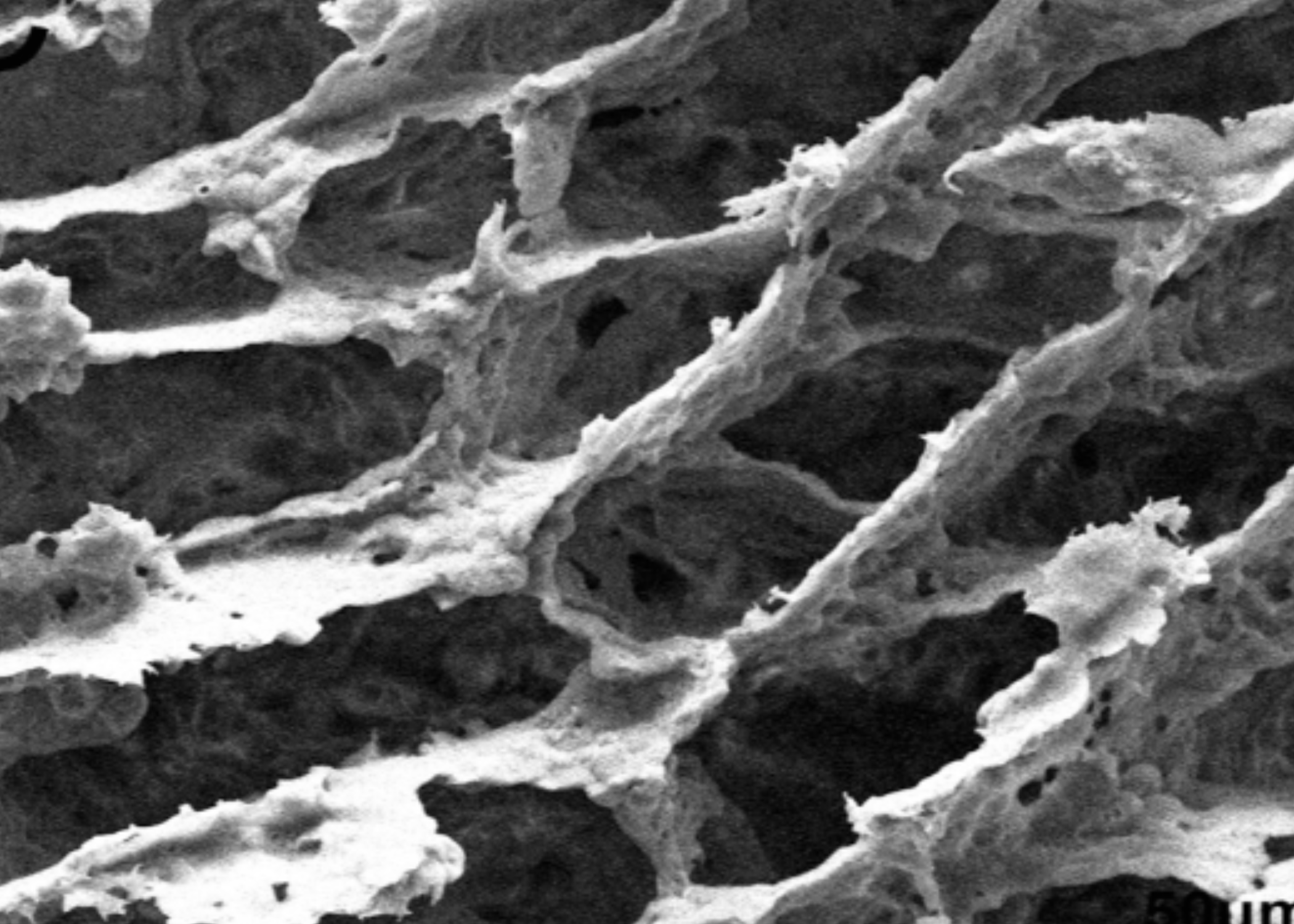
The scaffolds were manufactured using thermally induced phase separation, a technique that produces a porous three-dimensional structure without the use of solvents whose residues could pose a risk to surrounding tissue.
At 2, 4, and 8 weeks, the animals were evaluated using a range of complementary methods.
Micro-CT provided a quantitative three-dimensional picture of how much mineralized tissue had formed as a percentage of the total defect volume, expressed as BV/TV.
Hematoxylin and eosin histological staining revealed the overall tissue architecture under a light microscope. Safranin O staining identified areas of endochondral ossification, the process of bone formation through an intermediate cartilage stage that also plays a key role in the natural development of long bones.
Immunohistochemistry for type I collagen showed how actively cells were producing the main protein framework of the bone matrix. TRAP staining was used to identify osteoclasts, the cells responsible for bone resorption, whose excessive activity could potentially indicate an inflammatory or adverse tissue response.
What happened to the bone at each stage
The micro-CT results remained consistent throughout the observation period.
By the second week, the amount of newly formed bone in the hydroxyapatite-containing groups had already begun to noticeably exceed that observed with pure PLLA. This difference continued to increase through week eight.
There was, however, an important nuance. No statistically significant difference between HA/PLLA and PBLG-g-HA/PLLA was observed at any of the evaluation points. Both mineral-containing composites produced approximately similar results, even though surface modification was theoretically expected to improve particle distribution within the matrix.
Histology added important details.
In the HA-containing groups, abundant bone formation was observed, with a substantial proportion developing through the endochondral pathway, a mechanism that closely resembles physiological bone development.
Type I collagen staining showed the highest expression in the mineral-containing groups. This indicates more than simply the presence of calcium in the tissue. It reflects active maturation of the organic bone framework into which hydroxyapatite crystals can subsequently be incorporated.
The number of osteoclasts did not differ statistically among the four groups. Neither composite triggered excessive pathological resorption, suggesting that the materials did not provoke an abnormal tissue response in this model.
What this means for composite material engineering
The main conclusion of this study extends beyond the specific PBLG molecule.
It concerns the fundamental principle behind the design of bone implants: the presence of a calcium phosphate phase within a resorbable polymer matrix, rather than the polymer alone, can support osteoconduction, the ability of a material to provide a surface or structure along which new bone can grow.
A polymer without a mineral phase essentially remains a temporary mechanical scaffold. A polymer with a properly integrated mineral phase has the potential to “communicate” with bone cells in a chemical environment that is more familiar to them.
ResorBone™ does not use the specific PBLG surface-grafting technology described in this study, and none of the studies reviewed in this series directly tested ResorBone™ or biodrook products.
However, the principle underlying PBLG-g-HA/PLLA, combining a resorbable polymer with a calcium phosphate phase to support osteoconduction, is also fundamental to the material approach used in ResorBone™.
Each new study in this field, including this one, contributes to the broader scientific foundation behind this direction in bone biomaterials engineering and helps explain the rationale behind the material strategy selected for ResorBone™.
This article is a popular science review of a published study and does not constitute an advertising claim regarding the properties of a specific medical device. The conformity and clinical performance of biodrook implants are supported by the manufacturer’s own documentation.
Source: Liao L, Yang S, Miron RJ, Wei J, Zhang Y, Zhang M. Osteogenic Properties of PBLG-g-HA/PLLA Nanocomposites. PLoS ONE. 2014;9(9):e105876. doi:10.1371/journal.pone.0105876