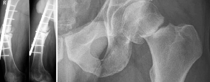
Pseudarthrosis, or false joint, is a condition in which bone fragments have failed to unite after a fracture within the clinically expected timeframe, and abnormal mobility develops at the site of injury. This complication leads to chronic pain, loss of limb function, and may result in disability. In this article, we will examine why pseudarthrosis occurs, how it is diagnosed, and which modern surgical approaches are used to restore bone union.
What is a false joint in simple terms?
A false joint is the result of impaired fracture healing. Normally, bone tissue is capable of regeneration: after a fracture, a bone callus forms between the fragments and gradually develops into mature bone. If this process stops or does not proceed properly, a gap remains between the fragments, and abnormal mobility develops at the fracture site — the so-called false joint.
Pseudarthrosis is diagnosed when a fracture fails to unite within the expected period of consolidation and the absence of healing progression is confirmed radiologically. This is not merely delayed union — it is a condition in which the biological conditions required for consolidation have been lost, and the likelihood of spontaneous union is low. Surgical intervention is generally required.
Potential consequences of pseudarthrosis:
- chronic pain at the fracture site;
- loss of weight-bearing function or limb strength;
- deformity and malalignment of the limb;
- risk of disability and the need for prolonged treatment;
- possibility of infectious complications.
Pseudarthrosis is not a congenital disorder (although there is a separate rare condition — congenital pseudarthrosis, which has a different etiology). This article discusses acquired pseudarthrosis resulting from a traumatic fracture.
How to recognize and confirm the diagnosis: symptoms and examinations
Pseudarthrosis is suspected when symptoms do not resolve within the clinically expected timeframe after a fracture and the patient’s condition does not improve.
Main symptoms:
- persistent pain at the fracture site that does not decrease over time;
- abnormal mobility at the site where the bone should have united;
- swelling, deformity, or a change in the shape of the limb;
- weakness, loss of function, inability to fully bear weight on the limb;
- absence of clinical and radiological progression of healing.
The diagnosis is established by an orthopedic trauma surgeon based on clinical examination and imaging findings.
Radiography in two projections is the primary diagnostic method. The images may show a gap between the bone fragments, sclerosis of the fragment ends (increased bone density), absence or insufficient formation of bone callus, deformity, or displacement of the fragments.
Computed tomography (CT) allows a more detailed assessment of the size and characteristics of the bone defect, the condition of the bone fragments and surrounding tissues, the presence of sclerosis or bone resorption, and the quality of previous fixation, if performed.
If infection is suspected (redness, wound discharge, fever, elevated inflammatory markers in blood tests), this is critically important for treatment planning. Infected pseudarthrosis requires a staged approach: first, removal of infected nonviable tissue and microbiological confirmation, followed by systemic antibiotic therapy, and only after the infection has been eliminated — definitive stabilization.
The final decision regarding treatment strategy is made by an orthopedic trauma surgeon after a comprehensive clinical and radiological assessment.
Why the bone does not heal: main causes and risk factors
Successful fracture healing requires four conditions: mechanical stability of the fragments, adequate blood supply, viable cells, and absence of infection. If even one of these conditions is impaired, the risk of pseudarthrosis increases.
Current reviews and AO Foundation materials emphasize that the risk of nonunion depends on fracture severity and the quality of restored stability, so these factors should be assessed together rather than in isolation.
Causes and risk factors can be divided into two groups: those related to the injury and treatment, and those related to the patient’s overall health.
Factors related to injury and treatment
Mechanical instability is one of the most common causes of pseudarthrosis. If the bone fragments are not adequately stabilized, continuous movement at the fracture site prevents bone callus formation. This may occur due to insufficient or inappropriate fixation, breakage or migration of fixation hardware, or premature weight-bearing on the limb.
Large bone defects or loss of a bone segment after injury create a gap that the body cannot fill on its own. The larger the defect, the greater the need for biological stimulation of bone union.
Infection (osteomyelitis) disrupts the normal healing process. Infected tissues cannot provide the conditions required for consolidation, so the infection site must first be debrided before stability can be restored.
An open fracture increases the risk of pseudarthrosis because of soft tissue damage, impaired blood supply, and possible infection.
More severe fracture morphology (comminuted, oblique, spiral) makes stable fixation more difficult to achieve and increases the risk of nonunion.
Factors related to the patient’s overall health
Smoking is a proven risk factor for pseudarthrosis. Nicotine impairs blood supply to bone tissue and reduces the activity of osteoblasts — the cells responsible for forming new bone.
Diabetes mellitus impairs microcirculation and slows regenerative processes. Patients with poorly controlled diabetes have a higher risk of nonunion.
Obesity is associated with an increased risk of pseudarthrosis, although the mechanism behind this association is not fully understood. It may be related to chronic inflammation, metabolic disturbances, and mechanical loading of the fracture site.
Impaired blood supply at the fracture site is a critical factor. If the bone does not receive sufficient oxygen and nutrients, the healing process stops.
A 2022 meta-analysis showed statistically significant associations between nonunion and smoking, diabetes mellitus, obesity, open fractures, and greater injury severity according to classification systems.
Types of pseudarthrosis and why they matter for treatment
Pseudarthroses are classified according to radiological and biological features. The most common classification divides them into two main types: hypertrophic and atrophic. Understanding the type of pseudarthrosis helps determine the appropriate treatment strategy.
Hypertrophic pseudarthrosis is characterized by excessive bone formation. Radiographs show pronounced callus formation around the fracture site, but the fragments have not united because of insufficient stability. The biological healing potential is preserved, but the mechanical conditions do not allow the consolidation process to be completed.
Treatment goal: provide stable fixation. Osteosynthesis alone is often sufficient without additional bone grafting because the biological capacity for union remains intact.
Atrophic pseudarthrosis is characterized by absent or minimal callus formation. The ends of the fragments are sclerotic, blood supply is impaired, and the biological healing potential is reduced or lost. It is often accompanied by a bone defect.
Treatment goal: not only stabilization, but also biological stimulation of union. This may include bone grafting, use of osteoconductive or osteoinductive materials, and restoration of blood supply.
| Characteristic |
Hypertrophic pseudarthrosis |
Atrophic pseudarthrosis |
| Radiological features |
Pronounced callus, “elephant foot” appearance |
Absence of callus, sclerosis of the fragment ends |
| Biological potential |
Preserved |
Reduced or lost |
| Main problem |
Fixation instability |
Poor blood supply, bone tissue deficiency |
| Treatment focus |
Stabilization |
Stabilization + biological stimulation |
The classification helps the surgeon determine whether improving fixation alone will be sufficient or whether biological stimulation of bone healing is also required.
Modern treatment approaches: how bone union is restored
Surgical treatment is the primary approach for established pseudarthrosis. Success depends on infection control (if present), achieving stability, and restoring a favorable biological environment.
Conservative methods (immobilization, physiotherapy) have limited effectiveness and do not eliminate the main causes of nonunion.
The general principle of pseudarthrosis treatment is based on three components:
- Elimination of infection (if present). Infected pseudarthrosis requires a staged approach: first, debridement of the infection site, removal of nonviable tissue, microbiological confirmation, systemic and, if necessary, local antibiotic therapy, followed by definitive stabilization.
- Restoration of mechanical stability. Bone union is impossible without stable fixation of the fragments.
- Provision of biological conditions for union. This may include bone grafting, the use of osteoconductive materials, and restoration of blood supply.
This concept is known as the “diamond concept” — a treatment framework that combines mechanical stability, vascularity, osteoinduction, osteogenesis, and osteoconduction with individualized selection of components. Understanding these principles helps individualize treatment strategy.
Surgical treatment: osteosynthesis and bone grafting
Osteosynthesis is the surgical fixation of bone fragments using metal implants. The choice of fixation method depends on the location of the pseudarthrosis, the type of fracture, and the condition of the bone:
- Plates and screws provide rigid fixation, particularly for metaphyseal fractures.
- Intramedullary fixation (nails) is commonly used for diaphyseal fractures of long bones.
- External fixation may be used for infected pseudarthrosis or complex defects.
The choice of a specific fixation system is made by the surgeon based on the clinical situation. There is no universally superior construct for all cases.
Bone grafting involves adding bone material to the pseudarthrosis site to stimulate union. The following are used:
- Autografts (the patient’s own bone, usually harvested from the iliac crest) — the “gold standard” because they contain living cells and growth factors. The disadvantage is the need for an additional surgical approach and the limited amount of available material.
- Allografts (donor bone) — do not contain living cells but provide osteoconduction (a scaffold for new bone growth). The risk of an immune reaction is minimal after processing.
- Synthetic osteoconductive materials (calcium phosphate ceramics, bioactive glass) — provide a scaffold for bone growth and are gradually resorbed. In a meta-analysis of nonunion treatment, synthetic substitutes demonstrated an overall union rate of approximately 90%, although outcomes vary depending on the material and study design.
Bone grafting is particularly important in atrophic pseudarthrosis and bone defects. In hypertrophic pseudarthrosis, improving fixation without additional biological stimulation is often sufficient.
Limitations of the method:
- Infected sites require debridement first.
- Large segmental defects may exceed the capabilities of standard bone grafting.
- Compromised blood supply or severe comorbidities reduce treatment effectiveness.
- Complex cases may require a staged or combined approach.
Patient-specific 3D-printed implants for bone defects
For significant bone defects, standard methods may be insufficient. In such cases, patient-specific 3D-printed implants may be considered.
Indications for patient-specific solutions:
- segmental defects or loss of bone tissue;
- complex anatomy where standard implants do not match the geometry of the defect;
- the need to restore limb length, alignment, or weight-bearing function;
- combining fixation with bone grafting or soft tissue reconstruction.
Advantages of customization:
Patient-specific design makes it possible to create an implant that precisely matches the anatomy of a particular patient. Based on CT or MRI data, a digital model of the defect is created, which is then used to manufacture an implant with the required geometry. This may improve biomechanical stability and simplify the surgical technique.
Reviews of patient-specific 3D implants report high rates of osseointegration and union in selected case series, alongside clinically significant complications; comparative studies with traditional methods are lacking. For critical hindfoot/ankle defects treated with 3D-printed titanium implants, an overall union rate of approximately 88% and a complication rate of ~17% have been reported, with infection and recurrent nonunion being the most common complications.
Limitations and requirements:
- A patient-specific implant must be designed with biomechanical loads and the clinical objective in mind.
- The implant material must meet regulatory requirements and be biocompatible with biological tissues.
- Active infection must be excluded before implantation.
- Patient-specific solutions do not eliminate the need for stable fixation and biological stimulation of bone union.
Role of biodrook:
biodrook specializes in the design and manufacture of personalized implants in collaboration with the surgeon. The process includes obtaining the patient’s medical data (CT, MRI), creating a digital model, approving the design, and manufacturing a sterile device ready to be provided to the physician. Patient-specific biodrook solutions may be considered for specific reconstructive tasks when standard approaches are limited, but the choice of treatment method always remains with the physician based on clinical assessment.
Recovery and prognosis: what to expect after surgery
After surgical treatment of pseudarthrosis, the patient needs time for bone consolidation and restoration of limb function.
Stages of recovery:
- Early postoperative period: pain management, wound care, and monitoring of soft tissue condition.
- Immobilization or weight-bearing restrictions as needed, depending on the location and type of fixation.
- Gradual mobilization under medical supervision to prevent contractures and maintain circulation.
- Follow-up radiographs to assess the progression of bone union.
Average healing times depend on the location and characteristics of the pseudarthrosis. In systematic reviews, the average time to union after surgical treatment of diaphyseal femoral nonunion was approximately 6–7 months, while for tibial nonunion it was approximately 7–12 months. Atrophic nonunion heals more slowly than hypertrophic nonunion.
These timeframes are averages. The specific recovery plan is individualized and depends on the type of pseudarthrosis, its location, the selected treatment method, comorbidities, and adherence to the physician’s recommendations.
Risks and complications:
- delayed union or recurrent nonunion;
- infectious complications;
- breakage or migration of fixation hardware;
- need for repeat surgical procedures.
To reduce risks, it is important to follow the weight-bearing regimen recommended by the physician, stop smoking, control blood glucose levels in diabetes mellitus, maintain adequate nutrition with sufficient protein and vitamin D intake, and attend regular follow-up appointments.
When to consult an orthopedic trauma surgeon
Timely consultation with an orthopedic trauma surgeon is critically important for successful treatment of pseudarthrosis.
Consult a physician if:
- pain and impaired limb function persist after a fracture beyond the expected healing period;
- there is no progress on follow-up radiographs;
- abnormal mobility develops at the fracture site;
- limb deformity develops;
- there are signs of infection: redness, swelling, wound discharge, fever.
After any procedure for pseudarthrosis, medical attention is also necessary if pain worsens, new symptoms develop, there are problems with the surgical wound or dressing, or complications are suspected.
Pseudarthrosis is a complex complication that requires an individualized approach. The earlier the diagnosis is established and treatment begins, the better the chances of restoring limb function. Adherence to the treatment plan and the physician’s recommendations are key factors for success.