
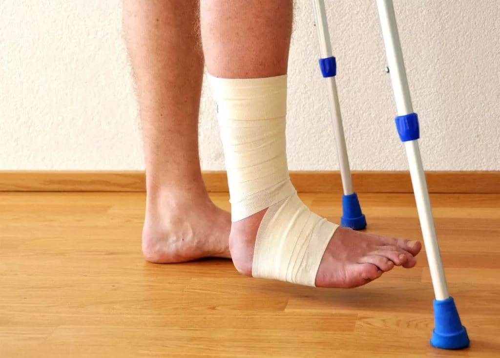

A gunshot fracture is a bone injury caused by the high-energy impact of a bullet or fragment, accompanied by destruction of bone tissue, wound contamination, and often significant soft tissue defects. Unlike ordinary fractures, such injuries require a specific staged approach: from emergency stabilization to prolonged reconstruction.
This article examines why gunshot fractures require a different treatment strategy, the stages a patient goes through, the fixation and bone defect reconstruction methods used today, as well as the role of modern technologies, including patient-specific 3D implants.
What is a gunshot fracture and why does it require a special approach?
A gunshot fracture occurs when a bullet or fragment penetrates the bone at high velocity. The impact energy damages not only bone tissue but also surrounding soft tissues, blood vessels, and nerves. The wound is always contaminated with particles of clothing, soil, bullet fragments, or shrapnel, creating a high risk of infection.
Key differences from ordinary fractures:
- High-energy injury. The bullet transfers significant kinetic energy to the bone, resulting in a comminuted fracture with a bone tissue defect.
- Wound contamination. A gunshot wound always contains foreign bodies and microorganisms, so the risk of infectious complications is significantly higher.
- Bone and soft tissue defects. Part of the bone tissue may be lost or nonviable. Soft tissues are also damaged, making wound coverage and healing more difficult.
- Combined injuries. Gunshot wounds are often accompanied by damage to blood vessels, nerves, tendons, or joints.
Because of these factors, gunshot fractures of the extremities are managed as open fractures: early intravenous antibiotics, debridement of nonviable tissues and bone fragments, and fracture stabilization are required. There is no universal protocol for all cases: the choice of treatment depends on the fracture location, extent of injury, condition of the soft tissues, and the patient’s overall condition.
Main stages of treatment: from stabilization to reconstruction
Treatment of a gunshot fracture follows a staged approach. The goal of the first stage is to save the patient’s life and limb and to control bleeding and infection. The goals of subsequent stages are to restore bone continuity, cover soft tissue defects, and restore limb function.
The rationale for the staged approach is that performing all tasks simultaneously is often impossible or unsafe. First, the patient’s condition and the fracture are stabilized, then infection is controlled, and only after that is definitive reconstruction undertaken. The individual treatment plan is developed according to the severity of the injury, associated injuries, and the capabilities of the medical facility.
Stage 1. Surgical debridement and temporary stabilization
The first stage of treatment begins with radical surgical debridement of the wound. All nonviable tissue, contamination, foreign bodies, and bone fragments that have lost their blood supply are removed. This is critically important for preventing infection.
After debridement, temporary fracture fixation is performed. External fixation devices are most commonly used, allowing the bone to be stabilized and the length and alignment of the limb to be maintained without requiring large incisions or introducing metal implants into a contaminated wound. External fixation also facilitates access to the wound for repeat debridement.
When indicated, fasciotomy is performed — surgical opening of fascial compartments to reduce soft tissue pressure and prevent compartment syndrome. If the soft tissue defect is extensive, the wound is not closed tightly; instead, early coverage with local or free flaps is planned.
For open fractures, early stabilization is recommended after thorough surgical debridement. In cases of substantial injury or when repeated revisions are necessary, temporary external fixation is preferred, while definitive internal osteosynthesis is performed after safe conditions for soft tissue coverage have been achieved. Definitive soft tissue coverage should be performed as early as possible; if this cannot be done during the initial operation, it should preferably be completed within 72 hours after injury.
Stage 2. Definitive fixation and soft tissue reconstruction
After infection has been controlled and the condition of the soft tissues has stabilized, definitive fracture fixation is performed. If the wound is clean, the soft tissues have been restored, and the fracture is stable, conversion to internal fixation with plates or intramedullary nails may be possible. In some cases, external fixation is maintained until complete union.
The timing and method of soft tissue defect coverage depend on the size and location of the defect. Small defects may heal by secondary intention or be closed with local flaps. Large defects require reconstruction with free flaps using microvascular anastomoses. Early and reliable soft tissue coverage reduces the risk of infection and creates favorable conditions for bone healing.
Reconstruction of the bone defect is often planned as a separate stage once infection has been controlled, the soft tissues have been restored, and the fracture has been stabilized. This may occur several weeks or months after the injury.
Staged treatment pathway for gunshot fractures
- Emergency care: bleeding control, immobilization, antibiotics
- Surgical debridement: removal of nonviable tissue
- Temporary stabilization: external fixation, fasciotomy when indicated
- Infection control: repeat revisions, early soft tissue coverage (preferably within 72 hours)
- Definitive fixation: internal osteosynthesis or prolonged external fixation
- Bone defect reconstruction: selection of method according to the clinical situation
- Rehabilitation: restoration of limb function
Sequence of gunshot fracture treatment stages: from initial debridement to reconstruction
Methods of fixation and bone defect replacement
The choice of fixation method and bone defect reconstruction strategy depends on the fracture location, condition of the soft tissues, infection risks, and the patient’s overall condition. Gunshot fractures often require a separate strategy for the bone defect because part of the bone may have been lost or removed during debridement.
Fracture fixation: external and internal — principles of selection
External fixation devices are appropriate during emergency stabilization when rapid fixation is required without additional trauma to the soft tissues. They allow control of limb length and alignment, provide access to the wound for repeat debridement, and do not require implantation of metal hardware into the contaminated area. Limitations of external fixation include patient discomfort, the risk of pin-site infection, and the need for regular care.
Internal osteosynthesis using plates or intramedullary nails is performed after infection has been controlled and soft tissue coverage has been restored. Internal fixation provides stability, allows earlier weight-bearing, and facilitates rehabilitation. Basic indications for internal osteosynthesis include a clean wound, stable soft tissue condition, and absence of active infection.
Factors that may require a change in strategy include the development of signs of infection, significant bone loss, fixation instability, or deterioration of the soft tissue condition. In such cases, external fixation may be resumed or definitive osteosynthesis may be postponed.
The choice between external and internal fixation depends on the severity of the injury, condition of the soft tissues, and time since injury; the approach is individualized according to the clinical situation.
Bone defect replacement: overview of approaches
When part of the bone is lost or removed, a bone defect develops and requires reconstruction. The main options include:
- Autografting. Transplantation of the patient’s own bone from another site (most commonly the iliac crest). An autograft has the best biocompatibility and osteogenic potential. Limitations include a limited amount of available material, an additional surgical procedure, and pain at the donor site.
- Allograft. Use of donor bone from a tissue bank. An allograft does not require an additional surgical procedure in the patient but has lower osteogenic potential and carries a risk of immunological reactions or infection transmission.
- Induced membrane (Masquelet technique). A two-stage technique: first, a cement spacer is placed in the defect, around which a biological membrane forms; the spacer is then removed and the defect is filled with an autograft. The method is suitable for large defects but requires two operations and prolonged treatment.
- Distraction osteogenesis. Gradual lengthening of the bone using an external fixation device. This method allows reconstruction of substantial defects without bone grafting but requires prolonged use of the device and strict adherence to the distraction protocol.
- Bone substitutes and biomaterials. Synthetic or biological materials that serve as a scaffold for new bone growth. They may be used alone or in combination with an autograft.
Factors considered when selecting a method include the size and location of the defect, infection status, condition of the soft tissues, the patient’s overall condition, resources of the medical facility, and the surgeon’s experience.
Systematic reviews show that autologous bone grafting, the Masquelet technique, and bone transport can provide high union rates in post-traumatic bone defects, but complication profiles and the strength of the evidence vary. High union rates have been reported for autologous bone grafting, although the included studies are heterogeneous. For bone transport, a meta-analysis of large diaphyseal defects showed relatively high union rates but higher rates of complications and repeat procedures compared with grafting techniques. A review of the Masquelet technique for segmental tibial defects indicates that union is usually achieved, often after additional procedures; however, complications are common and direct comparisons with other methods are limited.
| Method | Material/source | When considered | Limitations/risks |
|---|---|---|---|
| Autografting | Patient’s own bone | Small and medium-sized defects, absence of infection | Limited amount of material, donor-site pain, additional surgery |
| Allograft | Donor bone from a tissue bank | Large defects when autograft volume is insufficient | Lower osteogenic potential, risk of immunological reactions, risk of infection |
| Masquelet technique | Induced membrane + autograft | Large segmental defects | Two operations, prolonged treatment, high complication rate |
| Distraction osteogenesis | Patient’s own bone (lengthening) | Substantial defects when grafting is not possible | Prolonged use of the device, high complication rate, need for strict adherence to the regimen |
| Bone substitute materials | Synthetic or biological materials | Small defects, combination with autograft | Limited osteogenic potential, dependence on the specific material |
Methods of bone defect replacement: material source — advantages — limitations/risks
Modern technologies: patient-specific 3D implants in complex cases
For large or complex bone defects, multifragmentary areas, and situations requiring restoration of bone shape and load-bearing function, individualized solutions may be used. Patient-specific 3D implants are created according to the anatomy of a specific patient based on computed tomography data.
The materials used for such implants are most commonly titanium or biocompatible polymers. Titanium has high strength, biocompatibility, and osseointegrative properties. Bioresorbable materials can gradually dissolve in the body, but their use depends on the specific material and clinical objective.
Limitations of patient-specific implants include the need for multidisciplinary planning (surgeon, engineer, manufacturer), cost, manufacturing time, and the need for precise preoperative imaging. A patient-specific implant is not a universal solution for all bone defects but may be useful in complex cases when standard methods are insufficient.
When patient-specific implants may be appropriate
Patient-specific implants are considered in clinical situations where standard solutions are insufficient:
- large bone defects with complex geometry;
- multifragmentary fractures with bone loss;
- defects in anatomically complex regions (pelvis, scapula, proximal extremities);
- the need to restore load-bearing function and anatomical shape;
- inability to use an autograft because of insufficient quantity or the patient’s condition.
Prerequisites for the use of patient-specific implants include infection control, adequate soft tissue coverage, clearly defined reconstruction goals, and the ability to perform preoperative planning based on high-quality CT data. The decision to use a patient-specific implant is made by the surgeon after assessing the clinical situation.
How a patient-specific implant is created: from CT to implantation
The process of creating a patient-specific implant includes several stages:
- Data collection. The surgeon provides the manufacturer with the patient’s computed tomography data.
- Virtual planning. Engineers create a digital 3D model of the bone defect and design an implant that matches the patient’s anatomy. Medical modeling software is used.
- Surgeon approval. The implant model is reviewed and approved by the surgeon. Corrections are made if necessary.
- Manufacturing. The implant is produced using 3D printing on specialized equipment. Selective laser melting or other additive manufacturing technologies are used for titanium implants.
- Sterilization and quality control. The finished implant undergoes sterilization and quality control in accordance with medical standards.
- Intraoperative adaptation. During surgery, the surgeon places the implant and, if necessary, adapts it to the specific clinical situation.
Close collaboration between the manufacturer, surgeon, and medical facility is critically important for a successful outcome. Basic regulatory requirements for medical devices include certification, documentation, instructions for use, and quality control at all stages of production.
The official biodrook website states that patient-specific 3D implants are manufactured from CT/MRI data to fill bone defects after fractures, resections, and reconstructive procedures. The materials undergo preclinical testing in accordance with ISO 10993; the devices are manufactured and supplied sterile. The company reports certification of its quality management system according to DSTU EN ISO 13485:2018 in Ukraine and certification of its products in Ukraine.
From CT scan to placement of a patient-specific 3D implant
- Obtaining the patient’s CT data
- Creating a digital 3D model of the defect
- Designing the patient-specific implant
- Approval of the model by the surgeon
- 3D printing of the implant using specialized equipment
- Quality control and sterilization
- Delivery of the sterile implant to the surgeon
- Intraoperative implantation and adaptation
Workflow for manufacturing a patient-specific implant
Complications and prognosis: what occurs most often and how it is prevented
Gunshot fractures are associated with a high risk of complications. The main risks include:
- Infectious complications. Wound contamination, nonviable tissue, and the presence of foreign bodies create conditions for infection. Deep infection can lead to osteomyelitis — chronic inflammation of the bone that is difficult to treat.
- Nonunion or delayed union. Bone loss, impaired blood supply, infection, and unstable fixation can interfere with fracture healing.
- Loss of soft tissue volume and contractures. Significant soft tissue damage may lead to scar formation, restricted joint movement, and functional impairment.
- Pain syndromes. Chronic pain may result from nerve damage, scar formation, or bone instability.
Factors affecting prognosis include the extent of the initial injury, timeliness and radicality of debridement, stability of fixation, quality of soft tissue coverage, adherence to rehabilitation, the patient’s overall condition, and the presence of comorbidities.
Systematic reviews of gunshot fractures of long bones show that the risk of deep infection and nonunion exists, but the exact frequency varies considerably because of inconsistent definitions of complications and differences in treatment strategies. One review reports generally low infection rates in available case series but emphasizes the lack of standardized definitions and the limited ability to reliably determine the true incidence of deep infection. Complications are most commonly associated with vascular injuries, severe soft tissue damage, bone defects, comminuted or segmental fractures, and high-energy gunshot injuries.
Prevention within the surgical treatment strategy includes staged management, adequate debridement, stable fixation, early functional recovery, and early soft tissue coverage. According to the review, initial intravenous antibiotic therapy is recommended for all affected patients. It is important to understand that thorough debridement remains the standard for high-energy open gunshot injuries.
Rehabilitation: general principles of functional recovery
Rehabilitation after a gunshot fracture is a prolonged process that begins during inpatient treatment. The goals of rehabilitation are pain control, preservation of range of motion, gradual weight-bearing, prevention of contractures and muscle atrophy, and restoration of limb function.
The rehabilitation plan is individualized together with a physical therapist and depends on the fracture location, fixation method, condition of the soft tissues, and the patient’s overall condition. It is important to follow the weight-bearing regimen prescribed by the physician: premature loading may lead to displacement of fragments or fixation failure, while excessive restriction of movement may lead to contractures.
The patient is taught how to use the limb safely, perform exercises to preserve joint mobility, and gradually increase loading. Physical therapy includes passive and active exercises, massage, and physiotherapy procedures when indicated.
Key aspects of communication between the patient, physician, and rehabilitation specialist include regular examinations, radiographic monitoring, adjustment of the weight-bearing regimen according to the rate of bone healing, and discussion of pain and functional limitations. Specific recovery timelines depend on the clinical situation and cannot be determined in advance.
Conclusion
A gunshot fracture is a complex injury that requires a staged approach: from emergency stabilization and infection control to definitive reconstruction of the bone defect and restoration of limb function. The choice of fixation method and bone defect replacement depends on the fracture location, extent of injury, condition of the soft tissues, and the patient’s overall condition.
Modern technologies, including patient-specific 3D implants, expand reconstruction options in complex cases but are not a universal solution. The decision regarding the choice of treatment method is made by the surgeon after careful assessment of the clinical situation.