

Most scientific studies of bone implants are limited to weeks or, at best, a few months of observation. This is understandable: animal studies are expensive, time-consuming, and require careful ethical approval. But this creates an important gap in our knowledge. What actually happens to a resorbable material one year after implantation? Or a year and a half later? Does the composite lose contact with bone over such a long period, when the polymer phase is already actively degrading but the newly formed bone has not yet reached its final architecture?
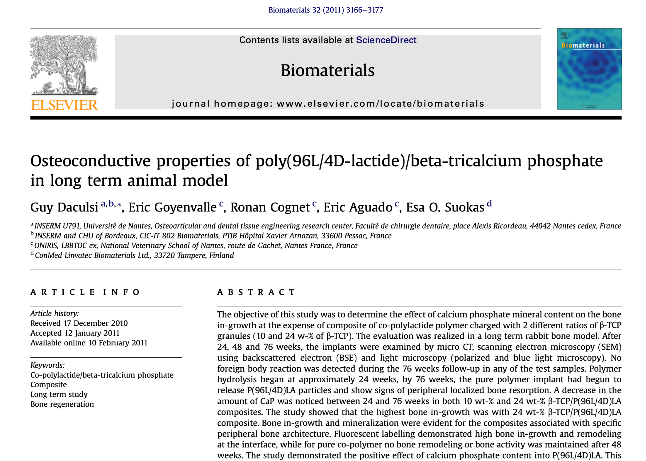
A French team led by Guy Daculsi from the University of Nantes set out to answer exactly these questions. Their publication in Biomaterials in 2011 reported an observation period of 76 weeks, almost a year and a half of continuous monitoring in a living animal model.
Three materials tested over the long term
The study compared three materials.
The first was pure P(96L/4D)LA copolymer, a polylactide containing L- and D-lactide monomers in a 96:4 ratio. The small proportion of D-lactide intentionally disrupts the highly ordered crystalline structure of pure poly-L-lactide, making the material less crystalline and allowing somewhat faster, more controlled degradation.
The second and third materials were composites based on the same polymer combined with β-tricalcium phosphate (β-TCP) at concentrations of 10% and 24%.
The materials were produced using twin extrusion followed by self-reinforcement, a process that aligns polymer molecular chains along the axis of the implant and significantly increases the mechanical strength of the final device.
Cylindrical specimens measuring 6 mm in diameter and 8 mm in length were implanted into critical defects of the lateral femoral condyle. In total, 54 cylinders were implanted in 27 rabbits, which were divided into observation groups of 24, 48, and 76 weeks.
The methodological depth of this study remains impressive even by modern standards.
The researchers used micro-CT for three-dimensional quantitative assessment, backscattered electron scanning microscopy for detailed analysis of the bone-implant interface, and polarized light microscopy with a special filter that made it possible to clearly distinguish oriented collagen in newly formed bone, soft tissue, and β-TCP particles.
One of the most sophisticated methods was fluorescent labeling of living animals using two tetracycline injections, administered 10 and 2 days before euthanasia. This effectively “highlighted” areas of active bone formation at a time point very close to the end of each observation period.
Diverging pathways: when hydrolysis becomes visible
During the first 24 weeks, all three groups behaved relatively similarly.
Hydrolysis of ester bonds within the polyester chains had only begun, and no major visible differences were observed.
Between weeks 24 and 76, however, the behavior of the three materials began to diverge significantly.
By week 76, the pure polymer group showed extensive degradation. The implant had begun releasing small particles directly into the surrounding tissue, and clear histological signs of localized resorption of the adjacent bone were observed.
In simple terms, the body had begun breaking down its own already formed peripheral bone more rapidly and less predictably than the polymer was creating controlled space for tissue replacement.
The two β-TCP-containing composites behaved very differently.
Direct bone contact did decrease somewhat over time, which is a natural consequence of material degradation. However, bone trabeculae remained attached perpendicular to the implant surface and maintained direct physical contact with calcium phosphate particles even when the surrounding polymer matrix was already undergoing substantial degradation.
At 76 weeks, bone contact with the composite containing 24% β-TCP was 64% higher than with the pure polymer.
Micro-CT also showed that three-dimensional loss of bone contact reached 54% in the pure polymer group at this time point, while it was substantially lower in both composite groups.
The most compelling evidence, however, came from fluorescent labeling.
Active bone remodeling at the implant-bone interface persisted throughout the entire observation period only in the β-TCP-containing groups.
In the pure polymer group, no active bone remodeling at the interface was detected after week 48. In the authors’ description, the interface had essentially become biologically inactive.
Buffering chemistry: why the mineral phase can reduce acidity
The mechanism can be explained by the basic chemistry of polyester degradation.
When an aliphatic polyester undergoes hydrolysis, water molecules attack the ester bonds within the polymer chains, generating carboxyl end groups and acidic degradation products, including lactic acid and related oligomers.
At sufficiently high local concentrations, these products can lower the pH of the surrounding tissue environment. This may irritate neighboring cells and contribute to increased or less predictable resorption, which is consistent with what was observed in the pure polymer group.
Calcium phosphate particles within the composite can act as an internal chemical buffer.
As they gradually release calcium- and phosphate-containing species, they can help neutralize acidic degradation products close to the site where those products are generated, reducing excessive deviation of the local pH from physiological conditions.
This provides an elegant solution to one of the fundamental challenges associated with resorbable polyester implants.
The broader principle of controlled degradation of a polymer matrix combined with an osteoconductive mineral phase is also relevant to the engineering approach behind ResorBone™, the material used to 3D print biodrook implants.
The study by Daculsi et al. investigated a specific P(96L/4D)LA/β-TCP system, not ResorBone™ or biodrook products directly. Nevertheless, it remains one of the longest and most methodologically detailed studies showing how this class of composites can behave in living bone tissue over a genuinely long healing period, not merely weeks, but many months and more than a year.
This article is a popular science review of a published study and does not constitute an advertising claim regarding the properties of a specific medical device. The conformity and clinical performance of biodrook implants are supported by the manufacturer’s own documentation.
Source: Daculsi G, Goyenvalle E, Cognet R, Aguado E, Suokas EO. Osteoconductive properties of poly(96L/4D-lactide)/beta-tricalcium phosphate in long term animal model. Biomaterials. 2011;32:3166–3177. doi:10.1016/j.biomaterials.2011.01.033