

A distal radius fracture, a fracture of the forearm bone close to the wrist, is statistically one of the most common fractures in the human body, accounting for approximately 8–17% of all bone fractures and up to 72% of all forearm fractures. In the United States alone, around 200,000 people sustain this injury each year. One of the most frequent and serious complications is malunion, when the bone heals in an incorrect, displaced position and disrupts the biomechanics of the entire wrist.
The standard surgical solution is a corrective osteotomy: the surgeon intentionally “re-breaks” the bone at the appropriate location and angle, then fixes it again using a titanium plate secured with screws. According to Japan’s Ministry of Health, Labour and Welfare, titanium is used in 95% of orthopedic implants and 70% of surgical implants overall.
But titanium is a compromise. It provides rigid fixation, leaving limited room for natural physiological flexibility, which may be particularly relevant in the developing skeleton. Documented, although relatively uncommon, issues include plate migration, temperature sensitivity, discomfort caused by palpable metal beneath the skin, and, in rare cases, allergic reactions. Perhaps most importantly, a second procedure is often required to remove the plate.
In a 2024 study published in 3D Printing in Medicine, a team from the University of Basel in Switzerland asked an ambitious question: could a titanium plate be replaced with a patient-specific, fully resorbable, 3D-printed wedge implant that gradually degrades after completing its mechanical function?
This question directly intersects with the broader engineering direction behind ResorBone™, the material used to 3D print biodrook implants.
A wedge instead of a plate: a different geometric philosophy
The idea behind the new approach is elegant in its simplicity.
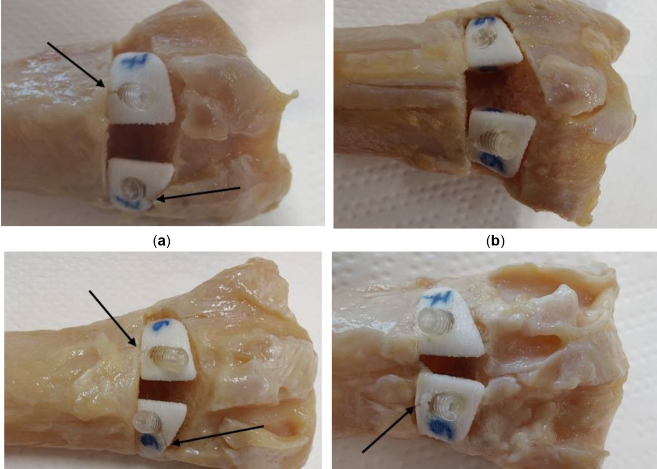
Instead of an external plate spanning the osteotomy site on the outside of the bone, the authors designed a compact wedge-shaped patient-specific implant, or WSPSI, that is inserted directly into the osteotomy gap, much like a wedge placed into a split.
The implant is fixed exclusively with resorbable compression screws, without any external metallic plate.
The process began with CT scanning of Thiel-embalmed cadaveric forearms, segmentation of the distal radius anatomy, and virtual osteotomy planning with an exact correction angle of 35 degrees, consistent with a previously published surgical reference protocol.
The osteotomies were incomplete, intentionally leaving the volar cortical layer intact. This technique provides additional natural stability even before the implant itself is inserted.
The wedge-shaped implants were designed with a clearance of only 0.5 mm from the dorsal bone surface to minimize the risk of material protruding beyond the natural bone contour.
Each wedge was designed to accommodate a 16 mm bioresorbable compression screw with a gradually increasing diameter from 3 to 3.7 mm.
The printing material was commercial Resomer® LR 706 S, a composite consisting of 70% PLDLLA and 30% β-tricalcium phosphate. It belongs to the same broad class of materials based on a resorbable polyester combined with a calcium phosphate phase that is also relevant to the engineering concept behind ResorBone™.
The implants were manufactured using Arburg Plastic Freeforming, a technology that combines aspects of injection molding with layer-by-layer additive manufacturing.
The moment of truth: axial compression until failure
Four cadaveric forearms received the new resorbable wedge implant, while a fifth was fixed using a conventional titanium plate as a direct reference.
Each specimen was embedded in acrylic resin, mounted in a universal testing machine, and loaded axially at a rate of 1 mm per minute until structural failure occurred.
The results were encouraging.
All PLDLLA/β-TCP wedge implants withstood at least 1,211 N of axial compression before any structural damage began.
This is an important figure. Previously published biomechanical data suggest that the peak impact force transmitted to the wrist during a typical forward fall from approximately shoulder height, around one meter above the ground, is about 1,021 ± 161 N.
In other words, even the weakest tested resorbable specimen tolerated a mechanical load above that associated with a realistic and clinically relevant fall scenario during the early postoperative period.
The best-performing specimen reached a maximum force of 2,736.4 N before cortical bone failure, while the titanium plate in the reference specimen failed at 2,415.6 N.
In its strongest configuration, the resorbable composite therefore demonstrated mechanical performance comparable to, and in this isolated specimen slightly higher than, the titanium reference.
The researchers also carefully analyzed how each sample failed.
In one specimen, a crack propagated outward from the edge of the drilled screw hole. This highlighted an area requiring further engineering optimization, for example by increasing the distance between the screw hole and the edge of the implant.
A material that does not contaminate itself during manufacturing
Alongside mechanical testing, the researchers carried out a separate degradation study.
Samples were tested in real time at 37°C and under accelerated conditions at 50°C for up to four weeks. Using the Hawkins accelerated-aging relationship, this was estimated to correspond to approximately 10 weeks of degradation at normal body temperature.
The results were cautiously but consistently encouraging.
The pH of the surrounding solution remained within a physiologically acceptable range of 7.4 ± 0.2 throughout the observation period, with no sharp acidification.
EDX analysis detected no metallic particles potentially introduced from the 3D printer nozzle during manufacturing. This was an important confirmation that the production process did not contaminate the composite with foreign metallic particles, despite the ceramic β-TCP phase being harder than the metal nozzle.
Sample mass loss during accelerated degradation was very small, ranging from 0.002% after 24 hours to a maximum of 0.467% at the longest accelerated-aging time point, suggesting a slow and controlled degradation process.
The study also revealed several important practical limitations, which the authors openly acknowledged.
The measured glass transition temperature of the material was 50.26°C, substantially lower than the manufacturer’s stated specification of 60°C. The authors associated this difference with the thermal and mechanical processing involved in 3D printing, during which the material was exposed to temperatures of approximately 205°C and pressures of around 200 MPa.
This is an important reminder that the manufacturing process itself can alter the physical properties of the original raw material and therefore has to be carefully controlled and validated at every stage.
Mechanics, chemistry, and what still needs to be studied
This study provides an important signal for the broader field of 3D-printed resorbable implants.
Patient-specific implants made from resorbable polyester/β-tricalcium phosphate composites can demonstrate mechanical strength in the same general range as titanium fixation while maintaining slow, controlled degradation.
This is also relevant to the broader engineering direction behind ResorBone™.
However, the material tested in this study was specifically Resomer® LR 706 S, not ResorBone™, and the experiments were conducted using prepared cadaveric forearms rather than living patients or biodrook devices.
The authors also acknowledge an important methodological limitation: the study evaluated axial mechanical loading and material degradation, but not the biological response.
In other words, the study did not investigate how living bone would heal in direct contact with such a wedge implant. That remains an essential next research step for this entire class of devices.
Even so, the mechanical and physicochemical findings strengthen the engineering rationale for exploring resorbable calcium phosphate composites as an alternative to permanent metallic fixation in selected trauma and reconstructive applications.
This article is a popular science review of a published study and does not constitute an advertising claim regarding the properties of a specific medical device. The conformity and clinical performance of biodrook implants are supported by the manufacturer’s own documentation.
Source: Jakimiuk A, Maintz M, Müller-Gerbl M, Thieringer FM, Keller M, Guebeli A, Honigmann P. 3D-printed patient-specific implants made of polylactide (PLDLLA) and β-tricalcium phosphate (β-TCP) for corrective osteotomies of the distal radius. 3D Print Med. 2024;10:42. doi:10.1186/s41205-024-00240-z