

There is an intuition about bone biomaterials that seems so obvious it hardly needs to be proven: if hydroxyapatite is the main mineral component of natural bone, then the more of it an implant contains, the better and faster the surrounding bone should heal.
This logic has shaped the development of calcium phosphate materials for decades. However, a study by a team from the University of Bergen in Norway and Alexandria University in Egypt, published in ACS Applied Materials & Interfaces in 2024, challenges this fundamental assumption. And it does so using an exceptionally comprehensive dataset spanning materials science, cell biology, and an in vivo animal model.
The real question, it turns out, is not “how much hydroxyapatite is in the material?” but rather “how quickly does that hydroxyapatite release calcium into the surrounding environment?”
These two parameters, mineral content and release rate, do not always correlate as simply as one might expect. Their relationship can critically influence whether a material actually supports bone healing or potentially inhibits it.
For any developer of 3D-printed calcium phosphate composites, including us as the team behind ResorBone™, this question is far from an academic abstraction. It directly informs how the balance between the polymer and mineral phases may affect the biological behavior of the material.
Four materials, printed using the same approach
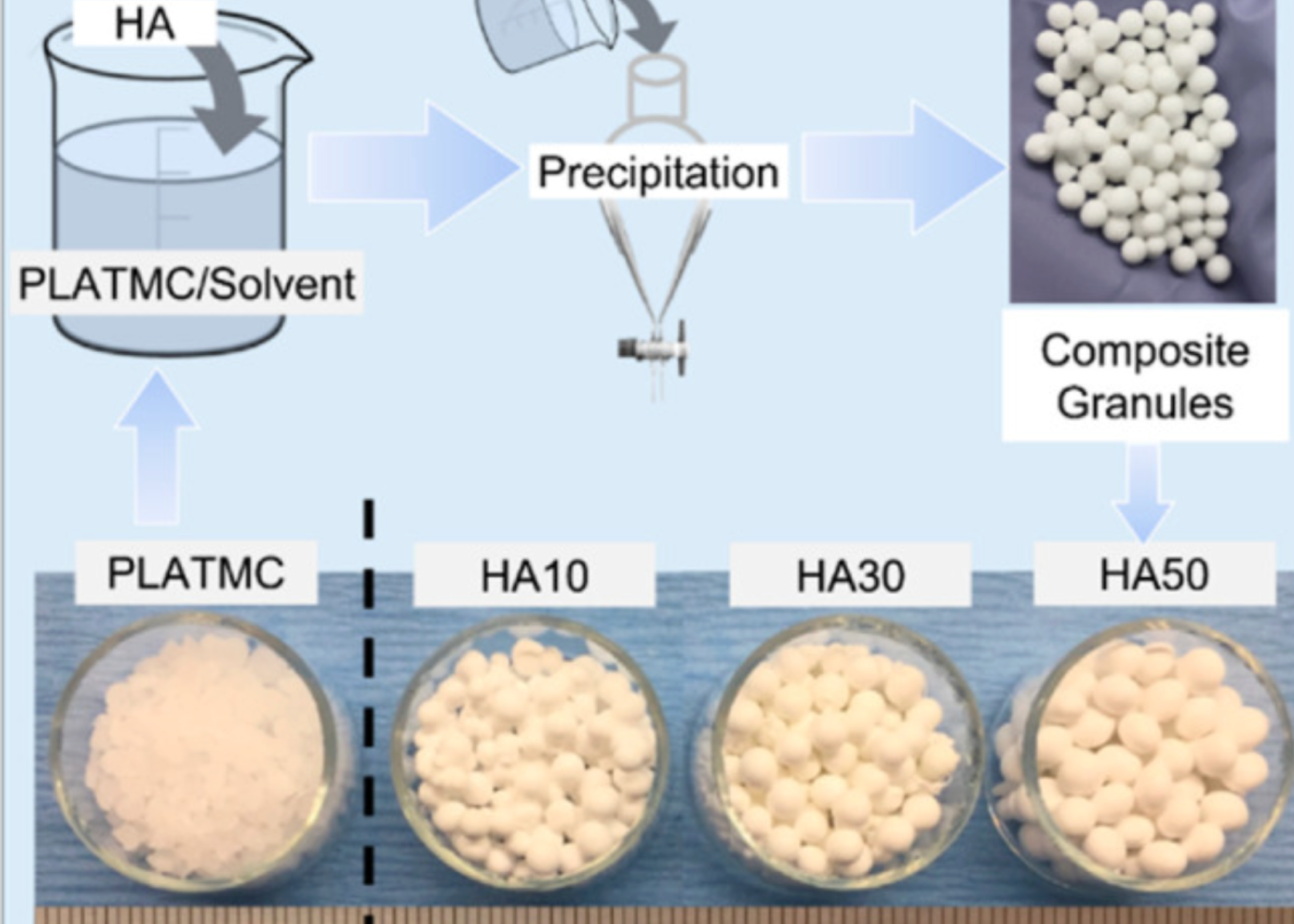
The researchers used poly(lactide-co-trimethylene carbonate) (PLATMC), a resorbable polyester with favorable mechanical properties and controlled degradation, as the polymer base.
It was combined with submicron hydroxyapatite particles, less than 200 nm in size, at three concentrations: 10%, 30%, and 50% by weight, creating the HA10, HA30, and HA50 groups. A pure PLATMC group served as the control.
All variants were manufactured using pneumatic extrusion 3D printing, part of the same broad class of additive manufacturing technologies used to produce modern three-dimensional medical implants, including the approach used to manufacture biodrook products.
Before biological testing, the researchers performed a comprehensive physicochemical characterization of each material, evaluating printability, surface roughness, wettability, mechanical properties, and thermal stability.
As HA concentration increased, surface roughness increased dramatically. In the HA50 group, it was almost eight times higher than in pure PLATMC or HA10.
At the same time, tensile strength gradually decreased, from 13.2 N/mm² for the pure polymer to just 7.9 N/mm² for HA50.
This alone highlights an important engineering consideration: adding a mineral filler requires balancing biological activity with the mechanical integrity of the final device.
But the real focus of the study was calcium release.
The researchers measured calcium release into a buffered solution for 100 days, an unusually long observation period for this type of test. This made it possible to evaluate not only an initial release burst but also the long-term dynamics of the material.
Two completely different calcium-release profiles
The HA10 group demonstrated slow, almost subtle release kinetics.
During the first 30 days, only around 27 μg of calcium per gram of material was released. Over the full 100-day period, cumulative release reached approximately 93 μg/g, corresponding to 4.8 μg/mL of solution.
These are relatively modest concentrations compared with what might typically be associated with an “active” calcium phosphate biomaterial.
HA30 and HA50 told a completely different story.
During the first two days, there was a sharp, almost explosive release of calcium, reaching 290 and 406 μg/g, respectively. Calcium release then continued at a consistently high level throughout the following 100 days without reaching a plateau.
Cumulative release reached 591–636 μg/g, more than six times the amount observed in the more conservative HA10 group.
According to the intuitive assumption that “more calcium means better mineralization,” these groups should have performed best.
But the opposite happened.
When cells say “too much”
In cultures of human mesenchymal stem cells seeded onto each material, the researchers observed a clear pattern.
By day 14, noticeably fewer viable cells had attached to HA30 and especially HA50 compared with PLATMC and HA10. The formation of mineralized extracellular matrix, assessed using Alizarin Red staining, was also lower in these groups.
By day 21, HA10 demonstrated a higher level of calcified matrix formation than all the other groups, including the pure polymer.
By day 28, the final evaluation point, pure PLATMC and HA10 showed a clear increase in biomineralization, while HA30 and HA50 demonstrated the statistically lowest levels.
EDX analysis of the material surfaces before and after 14 days of cell culture revealed another important detail.
In HA30 and HA50, surface calcium levels decreased significantly without evidence of new calcified matrix deposition.
In PLATMC and HA10, by contrast, surface calcium levels increased because cells were actively forming a new mineralized layer.
In other words, with moderate calcium release, calcium accumulated through cellular activity. With excessive release, calcium was simply being released from the material without producing the expected biological outcome.
The in vivo model confirmed what was seen in vitro
In a rabbit calvarial defect model, PLATMC and HA10 demonstrated the greatest amount of new bone formation at 4 and 8 weeks after implantation, with clear signs of contact osteogenesis, meaning newly formed tissue was directly apposed to the material surface.
HA30 and HA50, on the other hand, demonstrated what the authors described as distant osteogenesis. Bone formed away from the material, leaving a layer of fibrous tissue between the scaffold and newly formed bone, an indication of weaker osteoconductive behavior.
Based on the combined findings, the authors reached a specific and practically relevant conclusion: to preserve osteoconductive activity, cumulative calcium release should remain at approximately 25–30 μg per gram of material or below.
A lesson in dosage for ResorBone™ engineering
This study provides a valuable and somewhat counterintuitive reminder that working with biologically active materials can resemble drug dosing: the right amount can be beneficial, while an excessive amount may produce the opposite effect.
Bone cells are highly sensitive to the local concentration of extracellular calcium. A sudden, excessive influx of calcium ions may inhibit the very mineralization process that the mineral phase is intended to support.
Composite materials in the same broader class as ResorBone™, which is used to 3D print biodrook implants, are therefore not simply about maximizing the amount of hydroxyapatite or tricalcium phosphate in the composition. The engineering challenge is to achieve controlled, gradual degradation of the polymer matrix in a way that is compatible with the physiological pace of mineralization rather than competing with it.
The study by Hassan et al. investigated a specific PLATMC/HA system, not ResorBone™ or biodrook products directly. Nevertheless, the finding that there may be a “therapeutic window” for calcium release provides a valuable scientific reference point for the broader field of resorbable polymer-mineral composites.
This article is a popular science review of a published study and does not constitute an advertising claim regarding the properties of a specific medical device. The conformity and clinical performance of biodrook implants are supported by the manufacturer’s own documentation.
Source: Hassan MN, Eltawila AM, Mohamed-Ahmed S, Amin WM, Suliman S, Kandil S, Yassin MA, Mustafa K. Correlation between Ca Release and Osteoconduction by 3D-Printed Hydroxyapatite-Based Templates. ACS Appl Mater Interfaces. 2024;16:28056–28069. doi:10.1021/acsami.4c01472