

Sometimes, to truly understand why a modern product is designed the way it is, it is worth going back to some of the earliest peer-reviewed evidence in the field.
In 2008, a team of researchers from the Changchun Institute of Applied Chemistry of the Chinese Academy of Sciences published a study in Biomaterials that today reads almost like a classic textbook example. Precisely because of its methodological simplicity, thoroughness, and consistency, it remains worth examining closely, especially for us as a team developing ResorBone™ for 3D-printed biodrook implants.
The problem researchers were trying to solve more than 15 years ago
Poly(lactide-co-glycolide), better known as PLGA, is one of the most extensively studied resorbable polyesters in medicine, with a long history of clinical use and an established safety profile.
The problem was not its safety, but its biological “passivity.” On its own, without additional components, PLGA has no intrinsic osteogenic bioactivity. It does not actively stimulate osteoblasts to produce more new bone matrix. Instead, it mainly occupies space mechanically and gradually degrades.
Hydroxyapatite (HA), by contrast, is a structural and chemical analogue of the mineral component of natural bone. The calcium-to-phosphorus atomic ratio of the HA synthesized in this study, approximately 1.67, closely matches that of natural bone apatite.
However, raw, unmodified HA has an important drawback when used in composites: it tends to aggregate, forming clusters approximately 2–5 μm in size within the polymer matrix rather than distributing evenly across the pore surfaces of the scaffold, where the mineral is most useful for direct interaction with bone cells.
The researchers therefore proposed an elegant solution: synthesizing hydroxyapatite with poly(L-lactide) grafted onto its surface, creating a material they called g-HAP.
The idea was that a thin polymer layer would improve the compatibility of each HA particle with the surrounding PLGA matrix and prevent the formation of large aggregates. The grafted polymer accounted for approximately 5% of the particle mass, a relatively thin layer that nevertheless proved functionally meaningful in subsequent analyses.
Two independent tests: one in muscle and one in actual bone
The researchers evaluated three materials: pure PLGA, an unmodified HAP/PLGA composite, and the new g-HAP/PLGA composite.
They tested them using two parallel and complementary approaches.
The first involved intramuscular implantation in 18 rabbits, with three parallel samples of each material implanted in each animal. This model allowed the researchers to observe biodegradation and mineralization without the influence of mechanical loading or the natural bone microenvironment.
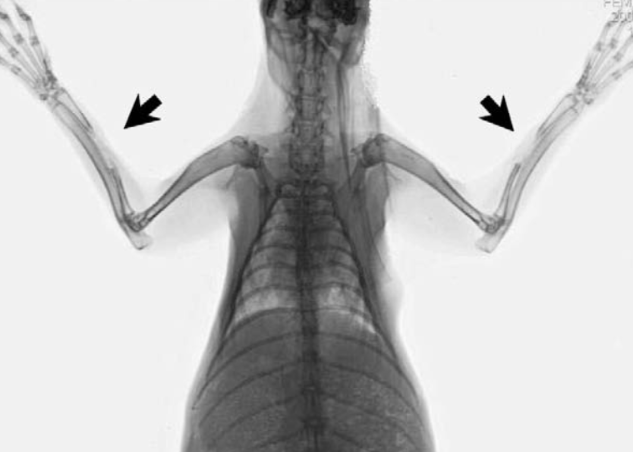
The second approach was considerably more complex and clinically relevant: reconstruction of a true critical-sized radial bone defect.
A 2 cm diaphyseal defect was surgically created in 12 rabbits. In this part of the experiment, the researchers also investigated the effect of the osteogenic factor BMP-2 by soaking g-HAP/PLGA scaffolds in a BMP-2 solution before implantation.
The implants in both parts of the study were evaluated at 4, 8, 12, and 20 weeks using a range of methods, including scanning electron microscopy, computed radiography, inductively coupled plasma atomic emission spectroscopy (ICP-AES) for precise quantitative measurement of calcium content, and conventional histology with hematoxylin and eosin and Masson’s trichrome staining.
Bone that formed like real bone
The results created a convincing picture at both levels of observation.
Following intramuscular implantation, both composite materials were substantially more stable than pure PLGA and demonstrated similar patterns of mineralization and biodegradation throughout the observation period.
Both composites showed two possible outcomes.
In some samples, extensive active mineralization occurred, accompanied by a sharp increase in calcium content, reaching as much as 18.7–19% of the material mass by week 20, almost six times the initial level.
Other samples of the same material underwent predominantly resorption.
This dual pattern appeared to depend on uncontrolled local factors in the microenvironment surrounding each individual specimen rather than solely on the chemistry of the material itself.
The more clinically relevant test, however, was the second part of the study involving true bone defects.
Here, the results were much clearer.
Hydroxyapatite-containing composites supported faster and more extensive new bone formation and demonstrated greater osteoconductivity than the pure polymer. The addition of BMP-2 to g-HAP/PLGA further accelerated this process.
By eight weeks, the calcium phosphate composite groups showed a genuinely integrated bone structure, with a clearly defined cortical layer and a fully developed medullary cavity.
In the pure PLGA group, the defect remained partially unfilled at the same time point, and bone formation occurred mainly from the peripheral periosteum rather than directly along the implant surface.
The comparison between modified and unmodified hydroxyapatite was also particularly interesting.
Surface modification of g-HAP did indeed produce a more uniform distribution of nanoparticles across the scaffold pore surfaces compared with unmodified HAP, where characteristic aggregation into 2–5 μm clusters was still observed.
However, the authors openly acknowledged an important point: despite this improvement in particle distribution, the final osteoconductivity and mineralization of the g-HAP/PLGA group were almost identical to those of the unmodified HAP/PLGA group, even though the actual hydroxyapatite content in g-HAP/PLGA was slightly lower.
This led to a practical engineering conclusion: the overall availability of calcium and phosphorus within the composite may be a key determinant of osteogenic activity, not merely the quality of particle dispersion.
An early but fundamental step toward the engineering logic behind ResorBone™
This study is one of the early and particularly illustrative examples in the scientific literature showing why combining a resorbable polyester with a calcium phosphate phase, rather than using the polymer alone, can provide osteoconductive properties in a bone implant.
In the more than 15 years since this work was published, numerous subsequent studies, including those discussed in previous articles in this series, have expanded and refined this fundamental concept across different animal models, polymer-to-mineral ratios, and manufacturing technologies, including modern 3D printing.
ResorBone™, the material used to manufacture biodrook implants by 3D printing, belongs to this broad family of polymer-mineral composites.
The 2008 study investigated a chemically related but distinct g-HAP/PLGA system developed by another research institution. It did not evaluate ResorBone™ or biodrook products.
Direct testing of biodrook products in animal models therefore remains a separate research stage, one that we regard as a logical and scientifically grounded continuation of this well-established research tradition.
Nevertheless, more than 15 years of accumulated evidence on resorbable polymer–hydroxyapatite composites, of which this study was one of the early systematic demonstrations in a true bone-healing model, provides an important scientific basis for the broader engineering approach applied in ResorBone™.
Source
Zhang P, Hong Z, Yu T, Chen X, Jing X. In vivo mineralization and osteogenesis of nanocomposite scaffold of poly(lactide-co-glycolide) and hydroxyapatite surface-grafted with poly(L-lactide). Biomaterials. 2009;30(1):58–70. doi:10.1016/j.biomaterials.2008.08.041
This article is a popular science review of a published study and does not constitute an advertising claim regarding the properties of a specific medical device. The conformity and clinical performance of biodrook implants are supported by the manufacturer’s own documentation.