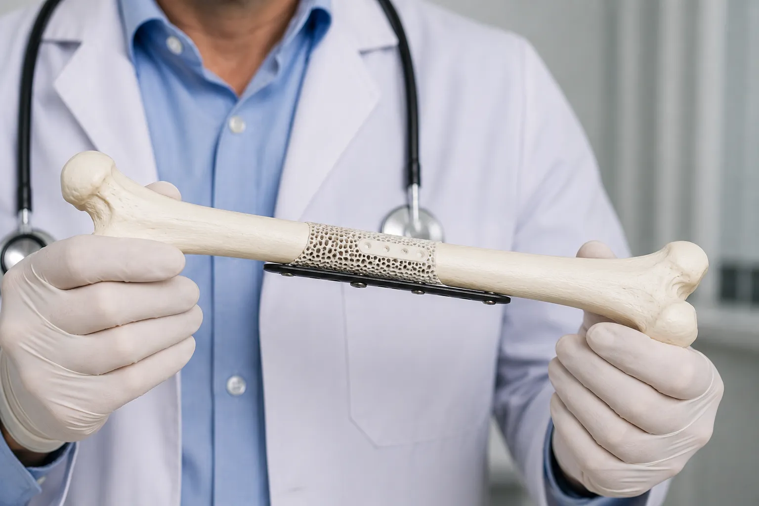
Artificial bone is a general term for synthetic materials used to restore bone tissue defects. They can replace the patient’s own bone or create conditions for its natural regeneration. In modern orthopedics, traumatology, and maxillofacial surgery, such materials are used in a variety of clinical situations — from post-traumatic defects to complex reconstructive procedures.
The choice of a specific material and method depends on the size of the defect, its location, the condition of the surrounding tissues, and the patient’s overall health. In this article, we will examine what artificial bone is, what types of materials are available, when patient-specific 3D implants are used, and what factors influence the physician’s decision.
What is “artificial bone” in orthopedics and surgery?
Artificial bone is not a single product but a group of bone substitute materials with different properties and forms. They are used for two main purposes:
- Defect replacement and structural support. The material fills a cavity or restores lost bone volume, providing mechanical support.
- Stimulation of the patient’s own bone growth. Some materials create a scaffold along which new bone can grow. This process is called osteoconduction — the ability of a material to serve as a structure along which new bone tissue forms.
ISASS guidelines describe bone substitute materials as products that can replace or supplement the patient’s own bone. Most categories of these materials primarily have osteoconductive properties and are used depending on the indications and characteristics of the specific product.
Artificial bone is not a universal solution. Different materials have different properties, and what is suitable for one clinical case may be ineffective in another.
When is bone tissue reconstruction needed: main indications
Bone tissue reconstruction may be indicated in various clinical situations:
- Post-traumatic defects. After severe fractures, especially open fractures or those involving loss of bone fragments.
- Consequences of tumor or cyst removal. The cavity left after surgical removal of pathologically altered tissue requires filling.
- Congenital or postoperative defects. Bone deformities that require correction and restoration of shape.
- Reconstruction in maxillofacial surgery. Restoration of jaw bone tissue, including preparation for placement of dental implants.
A systematic review and meta-analysis in the context of immediate implantation showed that the use of bone substitutes may reduce horizontal resorption without affecting implant failure rates.
Clinical guidelines for spinal surgery recommend considering bone substitutes in the presence of risk factors for nonunion or when the patient’s own bone is not optimal. Certain types of materials are acceptable only under specific conditions.
The decision to use bone substitutes is made individually. The physician evaluates the location of the defect, its size, the condition of the soft tissues, the blood supply to the area, and the patient’s overall health.
Artificial bone materials: bioresorbable and bioinert
Bone substitutes are classified according to their behavior in the body: materials that are resorbed and materials that remain as long-term structural support.
Any material must meet two basic requirements:
- Biocompatibility. The material must not cause a toxic or immune reaction.
- Suitability for the clinical task. The shape, volume, and mechanical properties of the material must be appropriate for the specific defect and load.
Bioresorbable materials: a scaffold for the growth of the patient’s own bone
Bioresorbable materials gradually resorb in the body and are replaced by the patient’s own bone tissue. They create a temporary osteoconductive scaffold — a structure along which new bone can grow.
Indications:
- small and medium-sized defects;
- areas with adequate blood supply;
- situations where natural bone repair and remodeling are expected.
Limitations:
Bioresorbable materials require adequate vascularization and stability of the area. If the blood supply is impaired or the defect is too large, natural replacement may be incomplete. The rate of resorption depends on the composition of the specific material and the conditions of its use.
A systematic review of non-spinal arthrodeses showed that the clinical effectiveness of different bone substitutes depends significantly on the specific material and indications, and for many products the level of evidence remains insufficient.
Bioinert materials: structural support without resorption
Bioinert materials are not resorbed in the body. They retain a stable shape and volume, providing long-term mechanical support. Depending on the material, direct bone contact — osseointegration (a direct structural connection between bone and the implant surface) — may occur, but replacement by the patient’s own bone does not take place.
Indications:
- large defects requiring permanent support;
- areas exposed to high mechanical loads;
- situations where restoration of shape is more important than biological integration.
Limitations:
Bioinert material remains in the body as long-term structural support. The decision to use it is made individually, taking into account long-term treatment goals and potential risks.
Forms of bone substitutes: from granules to patient-specific implants
Bone substitutes are available in different forms, each suited to specific clinical tasks.
Common forms:
- Granules or chips. Used to fill irregularly shaped cavities. The granules can be tightly packed to fill the defect.
- Pastes or gels. Convenient for injection using a syringe. Suitable for small defects or for filling complex anatomical areas.
- Blocks or plates. Used to restore bone contour and volume. A block can be shaped during surgery or manufactured in advance.
The form of the material corresponds to the clinical task: filling a cavity requires granules or paste, while restoring contour requires blocks or individually designed implants.
The need for precise restoration of anatomical shape has contributed to the development of patient-specific 3D solutions.
Patient-specific 3D implants: when they are appropriate and how they are created
Patient-specific 3D implants are personalized medical devices developed based on computed tomography or MRI data from a specific patient. They are designed to match the anatomy of the defect, helping restore its shape according to preoperative planning and improving the fit of the implant to the defect.
Indications:
- complex or large bone defects;
- the need for precise restoration of anatomical shape;
- reconstruction after tumor removal or trauma involving loss of a substantial amount of bone;
- cases where standard material forms do not meet the clinical requirements.
Various materials are used to manufacture patient-specific implants: titanium, medical-grade polymers, and bioresorbable composites. The choice of material depends on the location of the defect, mechanical load, and goals of the procedure.
Limitations:
Patient-specific implants require time to manufacture. The quality of the final device depends on the accuracy of the medical imaging and the thoroughness of planning. Such solutions are not universal for all types of defects — in some cases, standard materials may be sufficient or even preferable.
Stages of planning and manufacturing
The process of creating a patient-specific implant includes several sequential stages:
- Obtaining CT or MRI data. The physician provides medical images for analysis.
- Creating a 3D model of the defect. A digital model of the anatomical area is created based on imaging data.
- Virtual planning. The surgeon and engineer jointly agree on the shape, dimensions, and position of the implant.
- Manufacturing or printing. The implant is produced using specialized equipment and additive manufacturing technologies.
- Sterilization and preparation. The finished device undergoes the necessary procedures to ensure sterility.
- Surgical placement. The implant is provided to the surgeon for the procedure.
- Follow-up. Postoperative imaging is performed to assess the position of the implant.
This process requires interdisciplinary collaboration: the surgeon defines the clinical objective, the engineer develops the technical solution, and the manufacturer ensures production quality.
Example of a workflow with patient-specific implants
biodrook integrates the patient’s CT data, coordinates the implant design with the surgeon, and manufactures the device according to the patient’s individual anatomy. If necessary, surgical guides can be prepared for precise positioning of the implant during surgery.
The process includes a complete technological cycle: from digital modeling to the finished medical device.
Important: biodrook does not make clinical decisions. Indications for the use of a patient-specific implant, the choice of material, and the treatment strategy are determined by the physician after examining the patient.
Advantages and limitations: artificial bone vs the patient’s own bone (autograft)
An autograft — the patient’s own bone taken from another area of the body — is traditionally considered the reference option for bone grafting. ISASS recommendations emphasize that the patient’s own bone is the reference standard in spinal surgery, while substitutes are used as an alternative or adjunct, taking into account their mechanisms of action and limitations.
Autografts have limitations that make artificial materials a reasonable alternative in a number of clinical scenarios.
| Criterion |
Artificial bone materials |
Autograft |
| Additional surgical site |
Not required |
Required (donor site) |
| Volume limitations |
Depends on the manufacturer |
Limited available volume |
| Biological potential |
Depends on the material; osteoconduction is possible |
High; contains living cells |
| Predictability of shape |
High; a precise shape can be manufactured |
Limited; depends on the donor site |
| Risk of donor-site complications |
Absent |
Pain, infection, impaired function |
| Risk of surgical-site infection |
Depends on many factors; there is no risk of transmission of donor infections |
There is no risk of infection transmission from a donor; surgical infections are possible |
| Integration time |
Depends on the material and clinical situation |
May be faster; depends on the clinical situation |
Main conclusions:
- Autograft has high biological potential but requires additional surgical trauma and is limited in volume.
- Artificial materials make it possible to avoid a donor site, provide a predictable shape, and can be manufactured in the required volume.
- For large or complex defects, artificial materials, particularly patient-specific implants, may be one of the options depending on the clinical situation and indications.
A systematic review of non-spinal arthrodeses showed heterogeneous data: convincing results are available for some products, while evidence is insufficient for most. A meta-analysis in the context of immediate dental implantation demonstrated reduced horizontal resorption when substitute materials were used, with no difference in implant failure rates. Another systematic review found no statistically significant association between material type and implant stability.
There is no single gold standard for all cases. The choice between an autograft and artificial materials is a clinical decision that depends on the specific situation.
Possible risks and complications
The use of bone substitutes, like any surgical intervention, is associated with risks:
- surgical-site infection;
- incomplete defect replacement or nonunion;
- need for revision surgery;
- rare individual reaction to the material.
The frequency and profile of risks depend on the type of material, the clinical situation, the patient’s overall health, and adherence to surgical treatment protocols.
How does the physician choose the optimal material and method?
The choice of bone tissue reconstruction method is an individualized decision made by the physician after a comprehensive assessment of the clinical situation.
Key criteria:
- Size and location of the defect. Small defects may be filled with granules or pastes; large defects require blocks or patient-specific implants.
- Load on the area. Areas subjected to high mechanical loads require materials with appropriate strength.
- Age and comorbid conditions. The patient’s overall health, presence of chronic diseases, and blood supply influence the choice of method.
- Healing time. Some materials integrate faster, while others require a longer recovery period.
- Goals of the procedure. Restoration of function, aesthetics, or both.
- Availability of tissues and technologies. The possibility of harvesting an autograft and the availability of the necessary equipment and materials.
Clinical guidelines for spinal surgery recommend considering bone substitutes in the presence of risk factors for nonunion or when the patient’s own bone is unsuitable. Acceptability depends on the category of material and the clinical context.
Preoperative planning includes imaging (CT, MRI), assessment of the soft tissues, and discussion of possible treatment options with the patient. Complex cases may require consultation with several specialists.
Important: the decision to use a specific material or method is made by the physician after examination. If you have a bone tissue defect or are planning surgery, be sure to discuss all available options and their specific features with the appropriate surgeon.