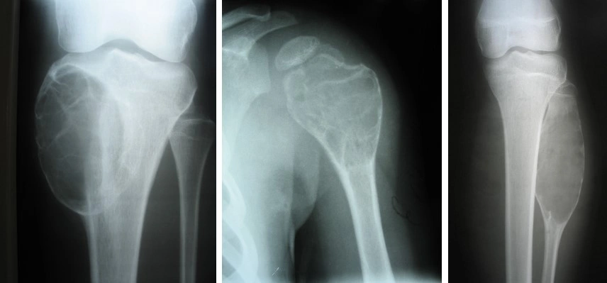
A bone cyst is a benign cavity within a bone filled with fluid. It is not a malignant tumor and does not spread to other organs. Bone cysts are most commonly found in children and adolescents, although they can occasionally occur in adults. This article discusses the nature of bone cysts, diagnostic methods, and general treatment approaches used by orthopedic trauma surgeons.
What is a bone cyst in simple terms?
A bone cyst is a fluid-filled cavity within a bone. It forms in bone tissue, does not contain malignant cells, and does not metastasize. It is a benign lesion that does not transform into a malignant tumor.
Bone cysts are most commonly found in children and adolescents aged 5 to 20 years. Typical locations include long bones: the femur, tibia, and humerus. Less commonly, cysts occur in the pelvic bones, spine, or other parts of the skeleton.
There are two main types of bone cysts:
- Unicameral (simple) bone cyst — the most common type; usually located in the metaphysis (the widened part of the bone near a joint), has a thin wall, and is filled with clear or yellowish fluid.
- Aneurysmal bone cyst — less common; contains blood-filled cavities separated by septa and may increase in size more rapidly.
The main potential danger of a bone cyst is weakening of the bone. The cavity thins the cortical layer (the dense outer shell of the bone), making the bone less resistant to mechanical stress and increasing the risk of fracture even with minor trauma.
Why do bone cysts develop and who is at risk?
The exact cause of bone cyst formation has not been definitively established. Several theories have been proposed to explain their development:
- Impaired local blood circulation in the bone. Local ischemia or venous obstruction is thought to contribute to cavity formation.
- Abnormalities of bone remodeling. During childhood, bones actively grow and remodel; disruption of this process may contribute to cyst formation.
- Microtrauma. Repeated minor injuries may affect bone structure and create conditions for cavity formation.
- Idiopathic cases. In many patients, no specific cause can be identified.
Who is affected more often:
- Children and adolescents — the main risk group; most cysts are diagnosed between 5 and 20 years of age.
- Boys — unicameral cysts are somewhat more common in boys than in girls.
- Adults — cysts may persist into adulthood or be detected for the first time in adults, but this is less common.
A bone cyst is not caused by infection, calcium deficiency, or poor nutrition. It is a localized structural abnormality of the bone that requires observation or treatment depending on the clinical situation.
Symptoms of a bone cyst and possible complications
A bone cyst is often asymptomatic. Many patients discover it incidentally — during radiographic examination for another reason or after an injury.
Possible symptoms:
- Aching pain with weight-bearing on the limb or after physical activity.
- Localized swelling in the area of the cyst if it is located close to the surface.
- Restricted movement or discomfort near a joint if the cyst is large.
- A feeling of weakness in the limb during prolonged physical activity.
Signs that require attention:
- Sudden pain after a minor blow or fall.
- Swelling and deformity of the limb.
- Inability to bear weight on the leg or move the arm.
These symptoms may indicate a pathological fracture — the most serious complication of a bone cyst.
Pathological fracture — the main danger of a cyst
A pathological fracture is a bone fracture that occurs after minimal trauma or even without trauma due to weakening of the bone structure.
How a cyst weakens the bone:
- The cavity within the bone reduces the thickness of the cortical layer — the dense outer shell of the bone.
- The larger the cyst and the thinner the wall, the lower the mechanical strength of the bone.
- Under load, the weakened area may fracture.
Typical situations involving a pathological fracture:
- A child falls while playing or participating in sports.
- An adolescent sustains a minor blow during everyday activity.
- A patient experiences sudden sharp pain during a normal movement without an obvious injury.
The risk of fracture depends on:
- The size of the cyst — larger cysts carry a greater risk.
- Location — cysts in the lower extremities (femur, tibia) have a higher risk because of constant weight-bearing.
- Thickness of the cortical layer — the thinner the wall, the higher the likelihood of fracture.
- The patient’s activity level — children and adolescents who participate in sports have an increased risk.
The risk of pathological fracture is the main factor influencing the choice between observation and surgical treatment.
How does a physician diagnose a bone cyst?
Diagnosis begins with a consultation with an orthopedic trauma surgeon. The physician takes a medical history and evaluates symptoms, the location of pain, and functional limitations.
Diagnostic sequence:
- Physical examination and medical history. The physician asks about symptoms, previous injuries, and the characteristics of the pain.
- Radiography — the primary method of initial diagnosis. The cyst appears as a radiolucent cavity with well-defined borders. Radiography makes it possible to assess its size, location, and cortical thickness.
- CT (computed tomography) — provides a more precise assessment of the cyst boundaries and cortical plate, detects occult fractures, and assists with surgical planning.
- MRI (magnetic resonance imaging) — confirms the cystic nature of the lesion, assesses the contents of the cavity, detects septa or fluid levels, and assists with differential diagnosis.
CT or MRI may be ordered if:
- Radiography does not provide a definitive answer.
- Other lesions need to be excluded.
- Surgical treatment is being planned.
- A pathological fracture or complication is suspected.
Differential diagnosis includes distinguishing the lesion from fibrous dysplasia, enchondroma, and giant cell tumor. In complex cases, a biopsy may be required.
Treatment: observation or surgery?
The choice of treatment strategy depends on the patient’s age, the size and location of the cyst, cortical thickness, presence of symptoms, fracture risk, and the patient’s activity level.
When observation is sufficient
Active surveillance is used for small asymptomatic cysts with sufficient cortical thickness and a low risk of fracture, particularly if the patient does not participate in contact sports or heavy physical labor.
Observation includes regular physician visits, scheduled radiographic or MRI examinations, activity restrictions, and informing the patient about symptoms that require immediate medical attention.
A more active treatment approach should be considered if the cyst increases in size, pain appears or worsens, the cortical layer becomes thinner, or a pathological fracture occurs.
When surgery is needed: curettage and bone grafting
Surgical treatment is considered for large cysts in weight-bearing areas, symptomatic lesions, a high risk of fracture, pathological fracture with instability, or progression of the cyst during observation.
In some cases, a simple cyst may partially or completely heal after a fracture with conservative management; the final decision is made individually by the physician.
Curettage is the primary surgical method and involves scraping out the contents and walls of the cyst. The resulting cavity may then be filled with bone grafting material to restore bone strength and support the remodeling process.
Bone grafting is the filling of the defect after cyst removal to provide mechanical support and stimulate new bone formation.
Materials used for bone grafting:
- Autologous bone — the patient’s own bone, usually harvested from the iliac crest; it has high biocompatibility and osteogenic potential but requires an additional incision and may cause pain at the donor site.
- Allogeneic bone — donor bone from a bone bank; it does not require an additional incision but has lower osteogenic activity compared with autologous bone.
- Synthetic biomaterials — artificial materials based on hydroxyapatite, tricalcium phosphate, or composites; they are biocompatible, gradually resorb, and are replaced by the patient’s own bone.
Some modern biocomposites can serve as a scaffold for new bone growth and gradually dissolve, creating space for the patient’s own bone.
Systematic reviews indicate that filling the defect after curettage is associated with favorable outcomes. Autologous bone may be associated with a lower recurrence rate compared with allogeneic cancellous bone or leaving the cavity unfilled. However, comparisons of different materials in terms of consolidation rates and complications remain limited and heterogeneous. It is not possible to reliably state that one material is superior to all others across all clinical outcomes.
Different bone grafting options are used in clinical practice according to indications and local protocols. biodrook develops biodegradable materials based on polylactic acid (PLA) with added hydroxyapatite, which gradually degrade in the body and, according to the manufacturer, support osseointegration. The choice of material depends on the clinical situation, the location of the defect, and the surgeon’s decision.
| Management approach |
Typical indications |
What it includes |
Goal |
Possible limitations |
| Observation |
Small asymptomatic cyst, low risk of fracture |
Follow-up visits, scheduled radiography/MRI, activity restrictions |
Avoid surgery if the cyst does not progress |
Requires patient compliance and regular monitoring; fracture risk remains |
| Surgery (curettage + bone grafting) |
Large cyst, symptoms, high risk of fracture, pathological fracture with instability |
Removal of cyst contents, filling the defect with bone grafting material |
Restore bone strength, reduce fracture risk, relieve symptoms |
Surgical intervention, possible complications (infection, cyst recurrence), need for rehabilitation |
Which physician should you consult and what should you do next?
A bone cyst is treated by an orthopedic trauma surgeon. For children, consultation with a pediatric orthopedic trauma surgeon experienced in pediatric bone disorders is recommended.
Seek immediate medical attention in the following situations:
- Sudden severe pain in a limb after minor trauma or without an obvious cause.
- Limb deformity, swelling, inability to bear weight on the leg or move the arm.
- Suspected pathological fracture.
What to bring to the appointment:
- Previous radiographs or CT/MRI reports, if available.
- A list of medications the patient is taking.
- A brief history of symptoms — when the pain started, whether there was an injury, and how the symptoms have changed.
After the diagnosis is established, it is important to follow the physician’s recommendations regarding activity restrictions, undergo regular follow-up examinations, not ignore new symptoms, and discuss the possibility of surgical treatment with the physician if there are indications.
If surgery is recommended, the treatment method, material used to fill the defect, expected recovery time, and postoperative restrictions should be clarified.
Conclusion
A bone cyst is a benign lesion most commonly found in children and adolescents. The main danger is weakening of the bone and the risk of pathological fracture. The choice between observation and surgical treatment depends on the size of the cyst, its location, symptoms, cortical thickness, and the patient’s activity level.
For small asymptomatic cysts with a low risk of fracture, active surveillance may be sufficient. For large cysts, significant fracture risk, or the presence of symptoms, surgical intervention — curettage followed by bone grafting — may be considered.
The final decision regarding treatment strategy is made by an orthopedic trauma surgeon after a complete examination and assessment of the clinical situation. If you or your child has been diagnosed with a bone cyst, consult an appropriate specialist for a detailed evaluation and selection of the optimal treatment approach.