

In biomaterials science, there are two fundamentally different levels of how “good” a material can be in relation to bone, and the distinction between them is easy to miss if you do not know what to look for.
The first, more familiar level is osteoconduction: the ability of a material to passively support bone growth along its surface, acting as a compatible scaffold along which cells can migrate and deposit new matrix.
The second, much rarer and potentially more clinically valuable level is osteoinduction: the ability of a material to initiate the formation of entirely new bone tissue without any added cells or growth factors, even in a location where bone would not normally form, for example, within skeletal muscle.
For a long time, the scientific literature largely considered osteoinduction to be an almost exclusive property of porous calcium phosphate ceramic materials, with internal porosity regarded as essential for this effect.
A study by a team from the University of Twente and the biotechnology company Xpand Biotechnology in the Netherlands, published in Biomatter in 2014, challenged this established assumption. Its findings are also relevant to the engineering of 3D-printed devices based on composite materials from the broader class to which ResorBone™ belongs.
The idea: a dense material with an artificially created “protected space”
The key engineering concept was to manufacture the composite by extrusion, a process that does not require organic solvents at any stage of production.
Residual solvents in a finished implant can be extremely difficult to remove completely, and even trace amounts may be harmful to surrounding tissues.
The composite consisted of 50% poly(D,L-lactide) and 50% custom-produced nanoscale hydroxyapatite, with particles approximately 309 nanometers long and 70 nanometers wide.
FTIR spectroscopy confirmed successful incorporation of the mineral phase into the polymer matrix. The composite spectrum showed clear phosphate and hydroxyl absorption bands characteristic of hydroxyapatite and absent from the spectrum of pure PLA.
The extrusion process produced a dense, completely nonporous material, essentially the opposite of the type of structure traditionally considered necessary for osteoinduction.
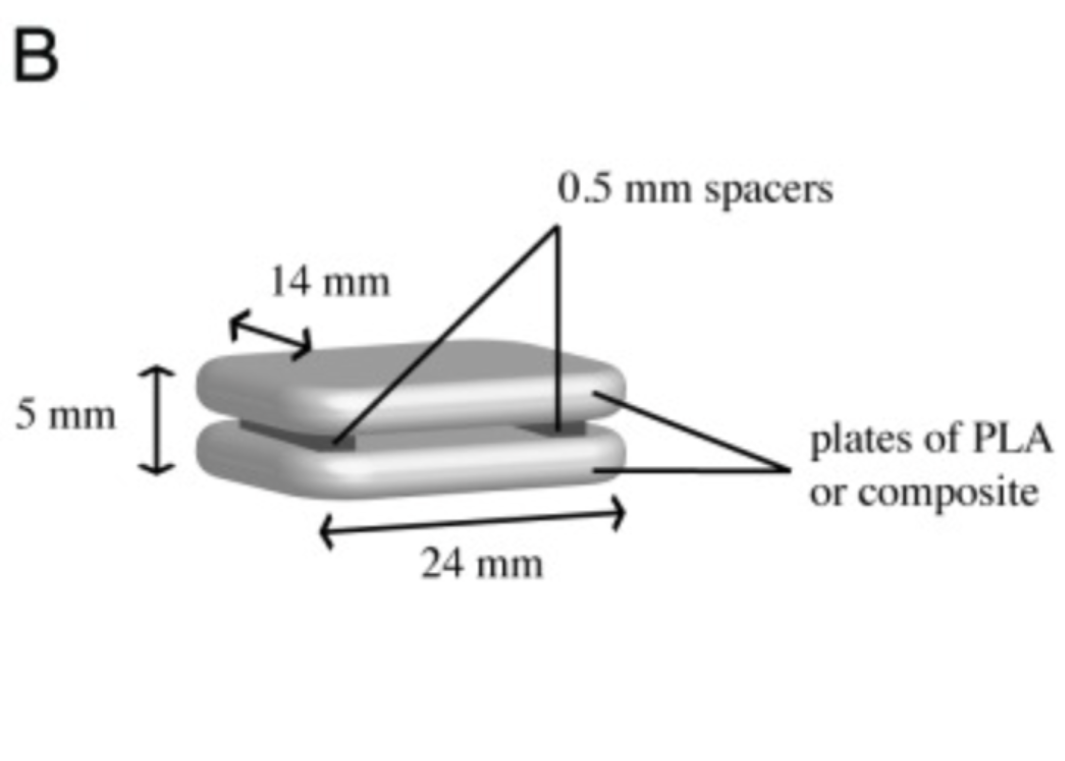
To determine whether such a dense material could nevertheless initiate bone formation, the researchers designed a somewhat unconventional but conceptually elegant implant.
Two dense composite plates were bonded together and separated by thin spacers. This created a narrow, artificially formed protected gap between the plates, effectively mimicking an internal pore even though the material itself remained completely nonporous.
Dogs, paravertebral muscle, and twelve weeks of observation
Twelve PLA/HA composite implants and five control implants made of pure PLA were implanted intramuscularly in eight adult mongrel dogs.
After 12 weeks, the animals were euthanized and the implants together with the surrounding tissues were collected for detailed histological analysis.
The results were already striking at the most basic stage, sample retrieval.
Of the five implanted pure PLA samples, only two could be located. At the sites where the remaining three should have been, the researchers found no detectable material or tissue reaction beyond normal muscle tissue. The polymer had apparently degraded without leaving any obvious trace.
By contrast, all twelve PLA/HA implants were successfully retrieved. Two were later excluded from histological analysis because of a technical issue during sample preparation.
The most important finding appeared under the microscope.
In all ten successfully analyzed PLA/HA samples, the researchers observed true heterotopic bone formation within muscle tissue, without the addition of cells or growth factors.
The greatest amount of bone formed near the entrance to the artificially created protected gap and in areas where degradation of the composite itself was most pronounced.
The newly formed bone had a physiological appearance, with mineralized matrix directly aligned with a layer of active osteoblasts. The maximum depth of bone ingrowth into the implant reached approximately 4 mm.
In neither of the two recovered pure PLA samples was any histological evidence of bone formation observed, despite clear signs of polymer degradation.
Cell biology supported what the animal model showed
Before the in vivo stage, the researchers also demonstrated that the PLA/HA composite steadily released calcium and phosphate ions into physiological saline throughout the entire 12-week observation period, without reaching a plateau.
Pure PLA, by contrast, released no measurable amounts of these ions over the same period.
Alkaline phosphatase, or ALP, activity in cells cultured on the composite was higher than on pure PLA in both tested media. The difference reached statistical significance on day 14 in the osteogenic culture medium.
What this means for 3D-printing geometry and architecture
The significance of this study extends beyond one specific PLA/HA composition.
It suggests that osteoinductive potential may also emerge in dense, completely nonporous composites based on a resorbable polyester combined with hydroxyapatite, provided that the overall implant design creates the right geometric conditions.
One particularly important factor may be the presence of a protected space where new bone tissue can develop and mature.
This is directly relevant to how engineers think about the geometry and internal architecture of 3D-printed implants made from composites in the broader ResorBone™ material class. The design of the implant itself, not only its chemical composition, may influence how actively the material environment supports new bone formation.
The material tested in this study was not ResorBone™ and the researchers did not evaluate biodrook products. It was a specially developed extruded PLA/HA composite with a 50/50 composition.
Direct testing of biodrook products in comparable animal models would therefore represent a separate future research step.
Nevertheless, this work expands the scientific evidence surrounding resorbable polyester-hydroxyapatite composites. It suggests that, under appropriately engineered conditions, such materials may do more than simply provide a passive surface for bone growth and may have the potential to contribute actively to the initiation of new bone formation.
This article is a popular science review of a published study and does not constitute an advertising claim regarding the properties of a specific medical device. The conformity and clinical performance of biodrook implants are supported by the manufacturer’s own documentation.
Source: Danoux CB, Barbieri D, Yuan H, de Bruijn JD, van Blitterswijk CA, Habibovic P. In vitro and in vivo bioactivity assessment of a polylactic acid/hydroxyapatite composite for bone regeneration. Biomatter. 2014;4:e27664. doi:10.4161/biom.27664