
Clinical Case 01
Personalized Bone Defect Reconstruction Resulting in Complete Functional Healing
Patient
38-year-old female
Injury
Fall from a height of approximately 2 meters
Diagnosis
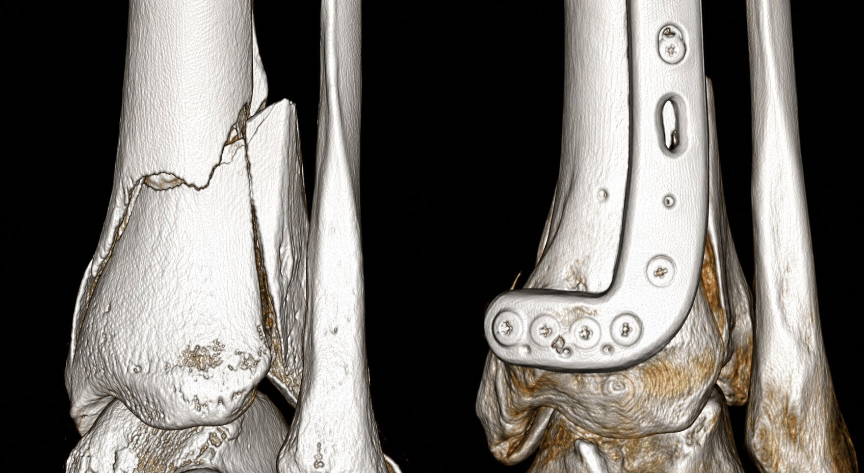
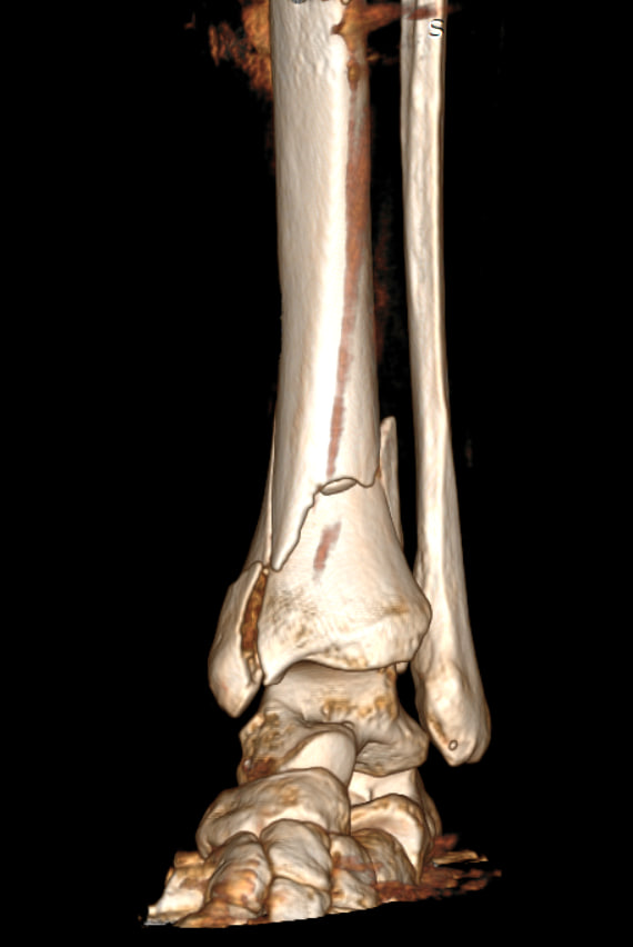
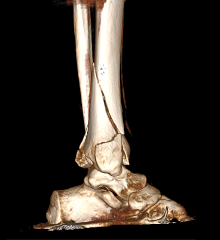
Comminuted intra-articular fracture of the distal tibial epimetaphysis (AO/OTA 44C)
Bone Defect
4.6 cm³
Clinical Challenge
Loss of subchondral bone support following reduction of impacted fracture fragments
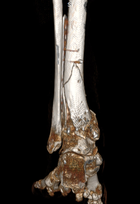
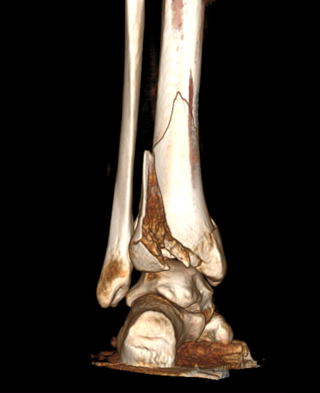
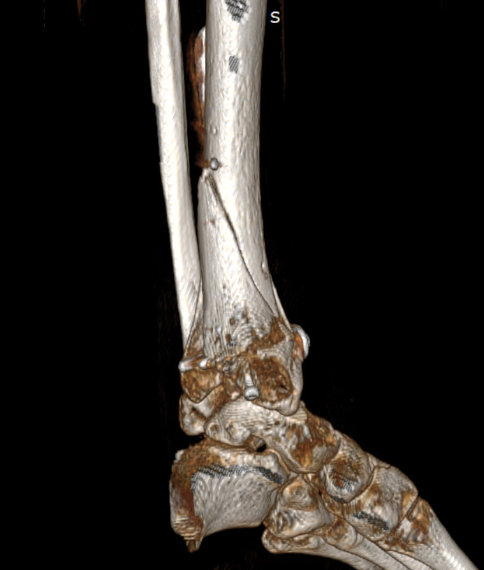
01 — Before Surgery
Before Surgery
A 38-year-old woman sustained a complex ankle fracture after falling from a height of approximately 2 meters. The injury resulted in significant bone loss beneath the joint surface, creating a defect that threatened long-term joint stability and function.
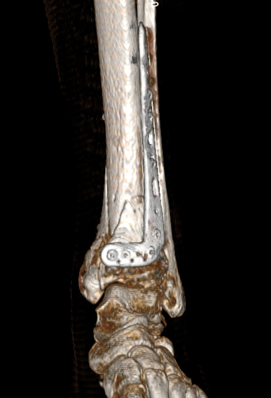
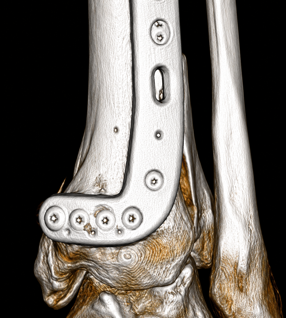
After emergency stabilization with an external fixation device, the patient underwent further evaluation by an orthopedic trauma surgeon. Once the surrounding soft tissues had recovered, a personalized treatment plan was developed to restore the bone defect and provide structural support using a custom 3D-printed bioresorbable ResorBone™ implant.
 BEFORE
 AFTER
AFTER
 BEFORE
 AFTER
AFTER
 BEFORE
 AFTER
AFTER
02 — Solution
What We Did
Following anatomical reduction of the fracture fragments, a complex bone defect measuring 4.6 cm³ remained beneath the joint surface. Using the patient’s CT data, the biodrook team developed a patient-specific 3D model and designed a custom ResorBone™ bioresorbable implant tailored to the exact geometry of the defect.
The implant was manufactured using 3D-printing technology and weighed only 6.4 g. Its precise fit provided structural support beneath the articular surface while creating favorable conditions for bone regeneration and long-term healing.
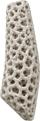
Custom ResorBone™ implant manufactured using 3D-printing technology. Weight — 6.4 g.
03 — Result
Results at 10 Months
Ten months after surgery, complete functional union of the affected segment was achieved. Follow-up CT imaging demonstrated advanced implant remodeling, with approximately 90% of the implant replaced by newly formed bone tissue.
The defect was almost entirely regenerated with the patient’s own bone, while dense integration between the implant and surrounding bone was observed without the formation of gaps or cavities. The reconstructed area demonstrated bone architecture and density closely resembling native bone. Residual implant material was visible only in small central regions.
- Complete functional bone healing
- Approximately 90% implant remodeling
- Near-complete replacement of the defect with new bone tissue
- Dense implant-to-bone integration without voids
- Restoration of bone structure closely resembling native bone
Videos
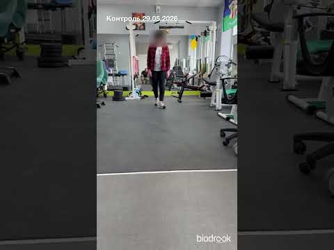
Watch the patient walking
Restored mobility after bone reconstruction
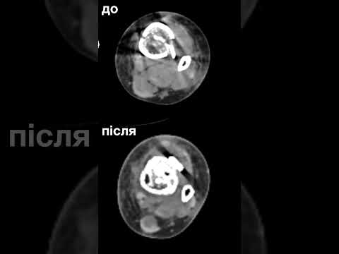
CT video before and after surgery
Computed tomography — comparative review
Patient Journey
Full story of treatment and recovery
HAVE A COMPLEX CLINICAL CASE?
Contact the biodrook team for a consultation. We will review your situation and propose a personalized solution.