
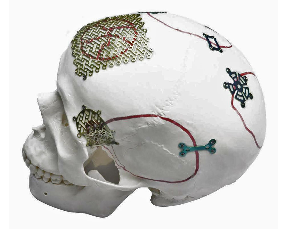

Cranioplasty is a surgical procedure to restore the integrity of the skull bones in the presence of a defect. A defect may result from trauma, tumor removal, an infectious process, or decompressive craniectomy. Restoring the bony coverage of the skull is important not only for protecting the brain but also for restoring normal anatomy and the patient’s appearance.
A patient-specific implant is a medical device created for a particular patient based on their CT data. This approach makes it possible to account for the anatomical characteristics of the defect at the design stage. In this article, we will examine how patient-specific implants differ from standard methods, when their use may be appropriate, what materials they are made from, and how the manufacturing process is carried out.
What is cranioplasty and what does “patient-specific implant” mean?
Cranioplasty is a surgical procedure to restore the integrity of the skull bones in defects of various origins. The aim of the intervention is to provide mechanical protection of the brain, restore normal cerebrospinal fluid circulation, correct the cosmetic defect, and improve the patient’s quality of life.
A patient-specific implant is manufactured according to the anatomy of a particular patient based on computed tomography data. The shape, thickness, and contours of the device correspond to the geometry of the defect and surrounding bony structures before surgery begins.
Unlike standard approaches, in which the surgeon shapes or adapts the material directly during the procedure, a patient-specific implant is created in advance using digital modeling and additive manufacturing technologies (3D printing). This makes it possible to anticipate the reconstruction outcome during the planning stage.
This article focuses on the technology used to create a patient-specific implant: planning, modeling, materials, and the manufacturing process. Surgical techniques for implant placement are beyond the scope of this material.
When is it appropriate to use a patient-specific implant?
A patient-specific implant may be preferred in certain clinical situations. The choice depends on the size and shape of the defect, its location, the condition of the soft tissues, and the clinical goals of reconstruction.
The use of a patient-specific implant may be appropriate in the following cases:
- Large and complex-shaped defects — when the geometry of the defect makes intraoperative modeling technically difficult or time-consuming.
- Cosmetically significant areas — the fronto-orbital region, temporal region, or parietal asymmetry, where accurate restoration of contours is aesthetically important.
- Need for high anatomical accuracy and symmetry — when it is important to restore the natural shape of the skull as precisely as possible.
- Repeat reconstructions — after previous unsuccessful attempts or complications, when a predictable outcome is required.
- Reduction of intraoperative modeling — in situations where shortening the duration of the procedure by using a ready-made device is desirable.
- Interdisciplinary cases — when a neurosurgeon and maxillofacial surgeon work together and a coordinated digital reconstruction plan is required.
Patient-specific implants are most commonly considered for large, irregular, or aesthetically critical defects, as well as for repeat reconstructions.
The final decision regarding the choice of method is made by the operating surgeon after assessing tissue condition, the patient’s overall health, and clinical factors.
How patient-specific implants differ from standard methods
Several approaches are used for cranioplasty. The choice of method depends on the size and location of the defect, the condition of the soft tissues, the clinical objective, and the surgeon’s experience.
Main methods:
- Patient-specific implant — manufactured from the CT data of a specific patient before surgery.
- Titanium mesh — shaped by the surgeon intraoperatively to close the defect.
- Bone cement (PMMA) — shaped directly during surgery.
Comparing these approaches helps to understand the clinical situations in which each may be appropriate.
| Criterion | Patient-specific implant | Titanium mesh | Bone cement (PMMA) |
|---|---|---|---|
| Anatomical accuracy | High; the shape corresponds to the patient’s anatomy based on CT data | Depends on intraoperative modeling by the surgeon | Depends on the shaping technique used during surgery |
| Need for intraoperative modeling | Minimal; the device is ready for implantation | Manual shaping is required during surgery | Material shaping and polymerization are required |
| Duration of surgery | May be shorter because the device is ready-made | Depends on the complexity of the defect and the surgeon’s experience | Depends on polymerization and modeling time |
| Predictability of the outcome | High; the shape can be assessed before surgery | Depends on the surgeon’s technique | Depends on the surgeon’s technique |
| CT/MRI visualization | Depends on the material (PEEK — minimal artifacts, titanium — moderate) | Moderate artifacts | Minimal artifacts |
| Aesthetics in cosmetically significant areas | High due to accurate reproduction of contours | Depends on the shaping technique | Depends on the shaping technique |
| Mechanical stability | High for both materials (PEEK, titanium) | High | Sufficient for most cases |
According to a 2026 systematic review, complication rates for PMMA did not exceed those of titanium implants in the analyzed studies. Some review publications describe fewer imaging artifacts and good clinical outcomes with patient-specific titanium implants.
The choice of cranioplasty method depends on the size and shape of the defect, the condition of the soft tissues, the presence of previous infection or failed reconstruction, and the patient’s cosmetic requirements.
The final decision is made by the physician based on the clinical objective, tissue condition, expected outcome, and available technologies.
Stages of creating a patient-specific implant: from CT images to the finished device
The process of manufacturing a patient-specific implant consists of several sequential stages, each of which is important for ensuring the accuracy of the device and its suitability for the clinical objective.
Main stages:
- Obtaining CT images and preparing DICOM data. The surgeon provides the patient’s computed tomography data in DICOM format (the standard format for medical images). The quality of the source data determines the accuracy of subsequent modeling. Important parameters include slice thickness, resolution, and anatomical coverage.
- Creating a virtual 3D model of the skull and defect. Based on the DICOM data, the engineer creates a digital three-dimensional model of the patient’s skull. The model makes it possible to accurately define the boundaries of the defect, bone thickness, and anatomical landmarks.
- Digital modeling of the implant by the engineer in collaboration with the surgeon; design approval. The engineer designs the implant based on the shape of the defect, skull symmetry, and the surgeon’s clinical requirements. The design is approved by the physician before manufacturing begins, allowing adjustments to be made during the planning stage.
- Manufacturing the device (3D printing or milling) from the selected material. After the design is approved, the implant is manufactured using additive technologies (3D printing, layer-by-layer production) or subtractive methods (milling, machining). The choice of technology depends on the material and device requirements.
- Post-processing, quality control, sterilization, and delivery to the clinic. The finished device undergoes verification of geometric accuracy, mechanical properties, and conformity with the approved design. After sterilization, the implant is delivered to the surgeon together with accompanying documentation.
How biodrook works with the physician during the manufacturing process
biodrook provides technological support at all stages of creating a patient-specific implant.
The collaboration process includes:
- Data exchange — the company provides the physician with requirements for CT data (format, resolution, slice thickness) to ensure the quality of the source information for modeling.
- Co-design with the surgeon — the engineer and surgeon jointly discuss the implant design and agree on its shape, thickness, fixation zones, and other clinically significant parameters.
- Approval protocols — before manufacturing, the surgeon receives the digital implant model for final confirmation.
- Device traceability — each implant is accompanied by documentation recording the stages of manufacturing, quality control, and sterilization.
biodrook reports DSTU EN ISO 13485:2018 certification, indicating the implementation of a quality management system for medical devices. On its product pages, the company also states that implants are tested for geometric accuracy and strength.
This approach allows the surgeon to monitor the process at key stages and receive a device that meets the clinical requirements of a specific patient.
Materials for patient-specific implants: PEEK and titanium
Two materials are most commonly used to manufacture patient-specific cranial implants: PEEK (polyether ether ketone) and medical-grade titanium. Each has properties that may be useful in certain clinical situations.
The choice of material depends on the clinical objective, defect location, imaging requirements, and other factors. There is no universally “best” material for all patients.
PEEK (polyether ether ketone)
PEEK is a biocompatible polymer used in craniofacial reconstruction because of its physical and biological properties.
Key properties:
- Elastic modulus close to that of bone — PEEK has an elastic modulus of approximately 3–4 GPa, which approaches that of cortical bone. This may contribute to more physiological load distribution.
- Radiolucency — PEEK is a radiolucent material and does not produce significant artifacts on CT. This facilitates postoperative monitoring and visualization of brain structures.
- Biocompatibility — the material is generally well tolerated by the body and does not cause pronounced inflammatory reactions in most cases.
- Potential for intraoperative modification — if necessary, a PEEK implant can be further adapted during surgery.
Typical clinical situations:
PEEK may be useful in cases where visualization of brain structures after surgery is important, as well as in patients who require regular CT or MRI monitoring. The material is also considered for reconstruction in cosmetically significant areas because of the ability to reproduce contours precisely.
Medical-grade titanium
Titanium is a metal widely used in orthopedics, traumatology, and craniofacial surgery because of its mechanical and biological properties.
Key properties:
- High strength and stability — titanium has an elastic modulus of approximately 110 GPa, providing high mechanical resistance to direct trauma.
- Proven biocompatibility — titanium has well-documented biocompatibility and a long history of clinical use.
- CT/MRI artifacts — titanium implants may produce moderate imaging artifacts, which should be taken into account when planning postoperative follow-up.
- Thermal conductivity — titanium has higher thermal conductivity than polymers; the clinical significance of this factor is limited for most patients and depends on the individual context.
- Fixation options — titanium implants can be fixed using screws or other fixation systems.
Typical clinical situations:
Titanium may be appropriate in cases where high mechanical stability is required, as well as in patients at risk of repeated trauma. The material is also considered for large defects where long-term structural strength is important.
The choice between PEEK and titanium depends on the clinical objective, defect location, imaging requirements, mechanical requirements, and other factors. The surgeon selects the material based on the patient’s individual characteristics and the goals of reconstruction.
Conclusion
Cranioplasty with a patient-specific implant is a method of restoring the integrity of the skull that makes it possible to account for the anatomy of a specific patient during the device design stage. Patient-specific implants may be appropriate for large or complex defects, cosmetically significant areas, and repeat reconstructions.
The choice between a patient-specific implant, titanium mesh, and bone cement depends on the clinical objective, tissue condition, the surgeon’s experience, and available technologies. Each method has its own advantages and limitations.
The process of creating a patient-specific implant includes obtaining CT data, digital modeling, design approval with the surgeon, manufacturing, and quality control. biodrook provides technological support at all stages and operates according to ISO 13485 quality management system standards.
If you are a physician considering the use of a patient-specific implant for a particular patient, you can contact the biodrook team to discuss the technical feasibility of manufacturing the device and the requirements for the source data.