
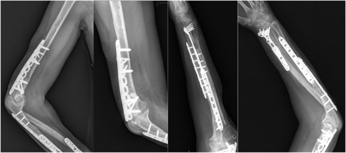

The Masquelet technique is a two-stage surgical method for treating large segmental bone defects based on the induced membrane phenomenon. The method is used for post-traumatic and post-infectious defects and complex nonunions of long bones when natural bone regeneration is insufficient. The main concept involves creating a biologically active membrane around a temporary spacer, which subsequently provides a favorable environment for bone grafting.
What is the Masquelet technique?
The Masquelet technique is a two-stage surgical method for treating large segmental bone defects. The method was developed for situations in which the amount of lost bone tissue exceeds the capacity for natural regeneration.
The basic principle is as follows: first, a temporary cement spacer is placed in the defect. Over several weeks, an induced membrane forms around it — a biologically active membrane with its own blood vessels and growth factors. During the second stage, the spacer is removed and a bone graft is implanted into the resulting “biological chamber.”
The technique is most commonly used to reconstruct defects of the long bones of the extremities — the femur, tibia, and humerus — after severe trauma, infectious processes, or surgical removal of affected tissue.
The outcome depends on many factors: the size and location of the defect, the condition of the soft tissues, blood supply, presence of infection, stability of bone fixation, and the patient’s overall health. The final decision regarding the choice of method is made by the surgeon after careful assessment of the specific case.
Principle of the method: the induced membrane and its role
The induced membrane is a biologically active membrane that forms around the cement spacer during the first stage of treatment. It performs several important functions that create a favorable microenvironment for subsequent bone formation.
Key functions of the induced membrane:
- Vascularization — the membrane contains numerous blood vessels that provide nourishment to the future bone graft.
- Barrier function — the membrane confines the defect space and prevents bone graft material from dispersing into the surrounding soft tissues.
- Local concentration of growth factors — the membrane produces biologically active substances that stimulate osteogenesis.
In clinical practice, the second stage is usually performed 6–12 weeks after the first, although the optimal interval depends on the location of the defect, the condition of the soft tissues, and the patient’s overall health. There is no single universal protocol suitable for all patients.
Stages of treatment using the Masquelet technique
The Masquelet technique consists of two distinct stages, each with its own clinical objectives. Both stages require surgical stabilization of the bone — internal or external fixation, depending on the location of the defect and the clinical situation.
First stage: debridement of the defect and placement of a cement spacer
During the first stage, thorough debridement is performed — removal of infected, necrotic, or nonviable tissue. This is a critically important step because the quality of debridement directly affects the treatment outcome.
After debridement, a temporary polymethyl methacrylate (PMMA) spacer — bone cement — is placed in the defect. The spacer fills the defect space, maintains bone length, and creates conditions for the formation of the induced membrane.
If infection is present or there is a high risk of infection, antibiotic-loaded PMMA may be used. The decision to incorporate antibiotics into the spacer is made individually. In systematic reviews of infected defects, the use of antibiotic-impregnated cement has been associated with a reduced need for revision procedures and more favorable bone healing outcomes.
At the same stage, stable fixation of the bone segment is ensured and soft tissue coverage of the defect is restored. Without stabilization and adequate soft tissue coverage, the formation of a fully functional membrane is not possible.
Second stage: bone grafting into the induced membrane
The second stage is performed after maturation of the induced membrane — usually 6–12 weeks after the first procedure.
The spacer is carefully removed while preserving the integrity of the induced membrane as much as possible. The membrane remains in place and forms a biological chamber into which bone graft material is implanted.
An autograft — the patient’s own bone, usually harvested from the iliac crest — is most commonly used. Autologous bone is considered the “gold standard” because of its osteogenic properties. In some cases, autologous bone may be combined with allograft (donor bone) or other bone substitute materials, but the choice of graft type is made individually.
After the defect is filled, stable bone fixation is maintained. Further treatment includes monitoring of bone union, rehabilitation, and gradual restoration of limb function.
Key prerequisites for success
Three prerequisites are critically important for achieving a favorable outcome:
- Infection control — radical debridement during the first stage and control of infection before bone grafting.
- Adequate soft tissue coverage — reliable closure of the defect during the first stage.
- Stable fixation — reliable stabilization of the bone segment throughout treatment.
When is the Masquelet technique used: main indications
The Masquelet technique is considered for reconstruction of large segmental defects of long bones in several major clinical scenarios.
Post-traumatic defects — the most common indication for the method. These occur after severe open fractures with loss of bone tissue when the size of the defect exceeds the capacity for natural regeneration.
Post-infectious defects and infected nonunions — situations in which a significant defect remains after surgical debridement of infected bone. The Masquelet technique makes it possible to first control the infection during the first stage and then perform reconstruction.
Defects after tumor resection — cases in which a segmental defect remains after removal of a benign or malignant bone tumor. The suitability of the method is assessed individually depending on the type of tumor and oncological prognosis.
Complex nonunions with bone loss — situations in which previous attempts to treat the nonunion have failed and the amount of lost bone is substantial.
In the literature, a critical-sized bone defect is most commonly defined as a defect longer than 2 cm or loss of more than 50% of the bone circumference. However, there is no single universal threshold, and assessment is always individualized.
The final decision to use the Masquelet technique is made by the surgeon after assessing the condition of the soft tissues, vascular status, infection control, and the ability to provide stable fixation.
Advantages, limitations, and possible complications of the method
The Masquelet technique has several clinically significant advantages, but it is also associated with limitations and risks that must be considered when selecting a reconstruction method.
Advantages
- Possibility of treatment in an infected environment — after thorough debridement, the method allows reconstruction even in post-infectious defects.
- Creation of a biological chamber — the induced membrane provides vascularization and local concentration of growth factors.
- Relative technical accessibility — unlike vascularized grafts, the method does not require microsurgical techniques.
- Compatibility with different fixation methods — both internal and external fixation can be used.
Limitations
- Two operations — the method requires two surgical procedures separated by several weeks.
- Prolonged treatment course — the total time from the beginning of treatment to complete bone union may take several months.
- Need for a sufficient volume of autograft — large defects may require a substantial amount of the patient’s own bone.
- Dependence on fixation stability — without reliable bone stabilization, the outcome is unpredictable.
- Dependence on soft tissue condition — adequate soft tissue coverage is critically important for success.
Possible complications
Systematic reviews show that complication rates with the Masquelet technique vary depending on the location of the defect and the clinical situation. According to different reviews, overall union rates range approximately from 77–96%, depending on location and the presence of infection.
For the lower extremity, some reviews have reported an overall complication rate of approximately 26%, with nonunion (approximately 9%) and deep infection (approximately 8%) being the most common complications.
The most common complications:
- Recurrence of infection — especially in post-infectious defects.
- Nonunion or delayed union — depends on the location and clinical situation.
- Graft resorption — partial resorption of the bone graft material.
- Donor-site problems — pain, infection, or other complications at the site of autologous bone harvesting.
- Fixation-related complications — problems involving metal implants or external fixation devices.
Factors that increase the risk of failure include postoperative infection of the bone graft, age over 65 years, insufficient stabilization, and inadequate debridement during the first stage.
Alternative methods
The Masquelet technique is not the only option for reconstructing large defects. Depending on the clinical situation, the following methods may be considered:
- Ilizarov bone transport — gradual movement of a bone segment to fill the defect.
- Vascularized bone grafts — transplantation of a bone segment together with its blood vessels.
- Combined methods — a combination of different approaches depending on the clinical objective.
The choice of method is always individualized and depends on the size of the defect, its location, the condition of the soft tissues, the presence of infection, and the experience of the surgical team.
The role of 3D printing: patient-specific spacers in the Masquelet technique
Traditionally, cement spacers for the Masquelet technique are shaped manually during surgery. The surgeon molds the PMMA spacer according to the shape and size of the defect. This approach is effective but has potential challenges: variability in shape, difficulty accurately reproducing the anatomy, and inconsistency in spacer volume.
Modern 3D-printing technologies create opportunities for producing patient-specific spacers or molds for shaping PMMA. Based on computed tomography data, an individualized spacer can be designed to more precisely match the anatomy of a specific defect.
Potential advantages of an individualized approach:
- More accurate reproduction of defect anatomy — the spacer can be designed according to the shape and dimensions of the missing bone segment.
- Standardized volume — the required amount of bone graft material for the second stage can be calculated in advance.
- Fixation planning — elements can be incorporated into the spacer design to improve interaction with fixation constructs.
However, it is important to understand the limitations of the evidence: clinical data on the advantages of 3D-printed patient-specific spacers in the Masquelet technique are currently limited. No reliable comparative studies have been identified in the available literature confirming that individualized 3D spacers improve clinical outcomes, reduce complication rates, or increase reconstruction accuracy compared with standard manually shaped PMMA spacers.
The decision to use individualized solutions is made by a multidisciplinary team, taking into account the specific clinical objective, technical capabilities, and safety requirements.
Companies working in the field of medical technology, including biodrook, are developing patient-specific 3D-printed solutions for orthopedics and traumatology. Biodrook manufactures patient-specific bone implants based on CT/MRI data, followed by sterilization and quality control.
What patients should know
If a physician recommends the Masquelet technique for reconstruction of a bone defect, it is useful to understand several key points.
Two-stage treatment — the method involves two operations separated by several weeks. The approximate interval between stages is usually 6–12 weeks, but the specific timing is determined by the surgeon depending on the clinical situation.
Stable bone fixation — throughout treatment, the bone must remain reliably stabilized. This may require the use of metal plates, rods, or external fixation devices. Weight-bearing restrictions on the limb remain in place until complete bone union is achieved.
Rehabilitation and monitoring — after the second stage, regular radiographic monitoring of bone union is necessary. Rehabilitation includes gradual restoration of movement and limb function under the supervision of a physiotherapist.
Infection control — in post-infectious defects, prolonged antibiotic therapy may be required. Adherence to recommendations for surgical wound care is critically important.
Smoking cessation — smoking impairs blood supply and slows bone healing. Smoking cessation significantly increases the chances of a successful outcome.
Individual differences — healing times may vary depending on the size of the defect, its location, the patient’s age, and overall health. In some cases, additional procedures may be required if complications or delayed union occur.
The final decision regarding the choice of treatment method is made by the surgeon after careful assessment of all factors. It is important to discuss expectations, possible risks, and alternative reconstruction options with the physician.