
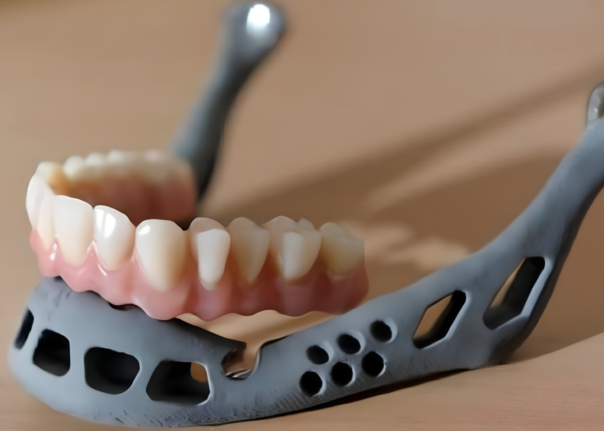

Most of the materials we discuss in this series are still making their way from the laboratory to the operating room. But clinical practice is already providing the first real examples of how resorbable 3D-printed scaffolds perform not in animal models, but in the body of an actual patient.
In January 2023, such a procedure was performed in Queensland, Australia, reportedly for the first time in the world using this particular combination of technique and material.
The patient, John Manwaring, had lost half of his lower jaw due to cancer. It was initially reconstructed using a conventional approach, with a segment of bone taken from his own fibula. However, following further cancer treatment, the graft began to deteriorate, and surgeon Michael Wagels had to look for another solution.
A scaffold instead of a piece of the patient’s own bone
The new approach used an implant developed by the Australian-Singaporean company Osteopore: a porous, biodegradable, 3D-printed scaffold wrapped in a thin layer of periosteum, the tissue covering bone that contains precursor cells capable of developing into osteoblasts.
The idea was not to immediately replace the missing bone with a ready-made bone segment, but to provide the body with a scaffold within which new bone could gradually grow.
According to Dr. Jing Lim, a representative of the company that developed the implant, the scaffold was specifically designed to allow tissue and blood vessels to grow not only around the structure, but throughout its entire volume.
This is a direct result of the porous architecture incorporated during the printing process, together with careful material selection for the specific clinical task.
Why vascular ingrowth matters more than shape alone
This technical detail is much more important than it may first appear.
Bone without an adequate blood supply cannot survive, regardless of how perfectly an implant reproduces the required geometry.
It is therefore not enough simply to fill a defect with a material of the correct shape. The internal architecture must provide blood vessels and cells with a physical pathway into the scaffold, rather than allowing growth only along its outer surface.
This is why porous three-dimensional architecture, rather than a solid block of material, remains a fundamental principle in the majority of modern bone scaffolds, including many of those discussed in previous studies in this series.
According to the treatment concept, the implant was expected to gradually resorb over approximately two years while the patient’s own mature bone tissue formed in its place.
This approach may eliminate the need for a second operation to remove the implant material, a consideration associated with permanent non-resorbable bone implants.
One clinical case and a broader scientific principle
This case illustrates a broader principle underlying the entire class of resorbable bone scaffolds: a porous structure capable of allowing blood vessels and cells to penetrate throughout its volume, combined with gradual and controlled material degradation synchronized with the formation of new bone.
This is also the broader engineering direction in which the class of composite materials that includes ResorBone™ is developing.
Clinical cases like this are particularly valuable because they demonstrate that the concept of a resorbable osteoconductive scaffold has moved beyond laboratory and animal studies and is already being applied in real surgical practice, including complex and unconventional clinical scenarios.
This article is a popular science review of published media coverage of a specific clinical case and does not constitute an advertising claim regarding the properties of any specific medical device. The case described involved a product manufactured by Osteopore, not ResorBone™ or biodrook products. The conformity and clinical performance of biodrook implants are supported by the manufacturer’s own documentation.
Source: Keane P. World-first Surgery to Help Man Regenerate Jaw Thanks to 3D Printing. 3D Printing. Published March 13, 2023.